NEW Images
These are our most recent images. They may belong to a new series of images, or expand one of the existing categories. If you are looking for previously unpublished material, this may be the best time to place your order. Please contact us for availability and pricing if you are interested.
«Your work is absolutely stunning and orders of magnitude better than most of what is out there (…)«
Dr. Jenny Whiting
Marketing & Business Development, Microscopy Australia, The University of Sydney.
Micronaut images are rights-managed. If you want to get a quote, please contact us, providing the following information: (1) image name, (2) specific use, (3) industry, (4) distribution area, (5) format, (6) circulation or print run, and (7) duration.
Please note that we cannot answer incomplete requests. Thank you.
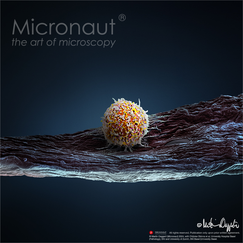
human T cell - engineered immune cell to fight cancer
Cellular Immunotherapy: this picture shows a human T cell from a immunocompetent humanized tumour model. Cancer immunotherapy with patients‘ own immune cells engineered to express chimeric antigen receptors (CARs) is a revolutionizing new treatment modality in hematologic oncology. CAR T cell therapy has achieved complete remission rates of 50 - 90% in patients with chemotherapy refractory or relapsed hematologic malignancies like diffuse large B cell lymphoma (DLBCL). By comparison, in DLBCL for example, the complete response rate to conventional therapies in patients with refractory or relapsed disease is below 10%. Still, up to one third of patients treated with CAR T cells relapse within a year after an initial complete response, there is a high burden of therapy associated toxicities and non-hematologic cancers have thus far not been amenable to redirected cellular immunotherapy. Apart from T cells, natural killer (NK) cells are a second cytotoxic lymphocytic lineage that have not been sufficiently explored clinically in the context of CAR therapy. Indeed, NK cells might be a promising alternative to T cells due to an inherent anti-cancer activity and a favorable toxicity profile. With a longstanding interest in human NK cells, our research includes engineering these cells as vehicles for redirected cellular immunotherapy. As a preclinical cancer model, we are using human immune system (HIS) mice carrying EBV-induced lymphomas. This allows the exploration of an autologous human cancer-immune system interface and its manipulation. We are specifically interested in engineering cytotoxic cells beyond redirection to enhance anti-cancer activity of the infused cell product. Another interest in the lab is functional analysis of genes important for human NK cell biology in vivo using HIS mice via genome editing approaches in the context of viral infection and homeostasis. These studies are designed to contribute to novel insights and improvements in the therapeuti
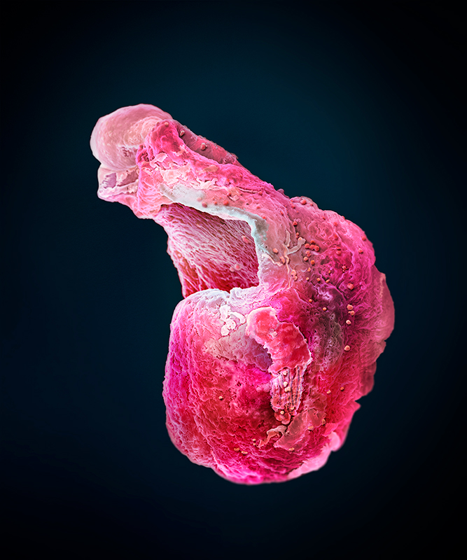
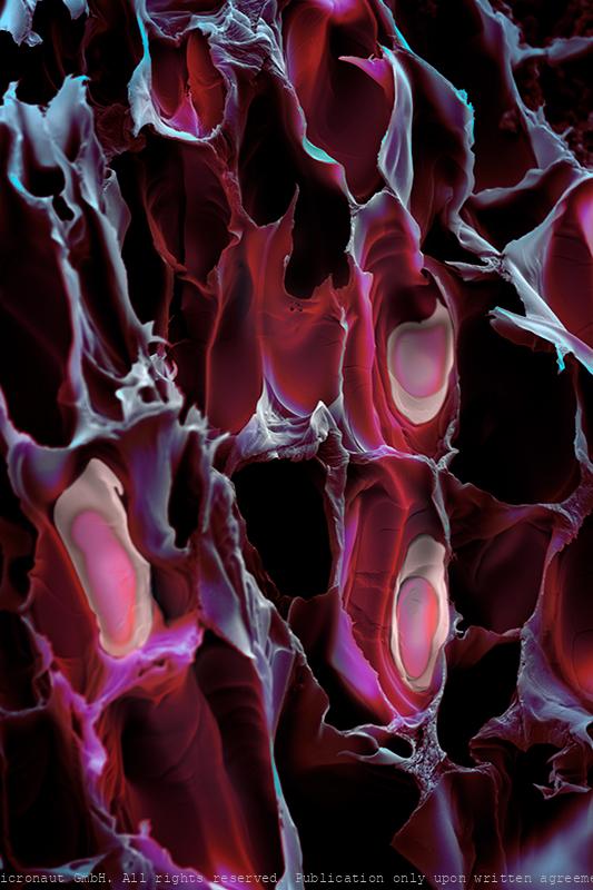
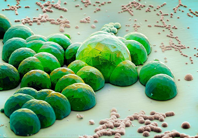
Synthetic mini hearts beat like the real ting!
Advancements in the development of patented synthetic human-like hearts allow researchers to study the development of the human heart and related disease on highly accurate models. This facilitates the development of innovative therapies and new pharmaceutical drugs to treat heart-related problems such as cardiovascular and metabolic diseases. Hand-colored scanning electron micrograph (SEM) by Martin Oeggerli in collaboration with Makoto Hara and Lucas Goguen of the Novartis Institute for BioMedical Research (NIBR). Annually an estimated 21 million deaths are related to disorders of the heart and blood vessels, and that number is growing. By creating synthetic organoids that beat like the real thing, scientists are unravelling the intricacies of the heart. Similar in size and development to fetal human hearts, these mini heart organoids are becoming increasingly complex and realistic.
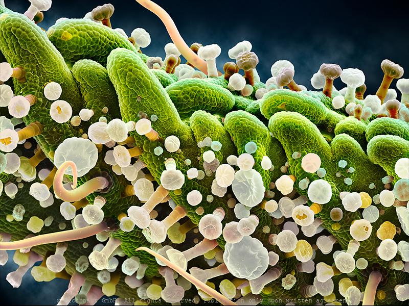
Wonderland Forest - The Walnut Leaf (Juglans sp.)
Under the powerful magnification of a scanning electron microscope (SEM), the leaf of a Walnut (XY sp.) looks like a forest location in Wonderland. And much like the forest in Wonderland it is so huge that it never seems to end for its many bizarre micro-organisms that tend to get lost there. And should it end at one point - at the edge of the leaf - it will continue on the next leaf, et cetera. If you look close, whatsoever, this micro-forest consists of glands and leaf hairs (trichomes) rather than bushes and trees. Glandular trichomes are multicellular secretory structures on the leaf surface which are able to synthesize and store various chemical compounds in order to protect the plant (leaf) from herbivorous insects, fungi, pathogens, extensive light, high temperature, and even UVB radiation. In Juglans regia you can find thousnads of these especially on the emerging (young) leafs. Many of these chemicals are medically relevant and thus, Junglans regia has been widely used in traditional medicine for very different ailments including: helminthiasis, diarrhea, sinusitis, stomachache, arthritis, asthma, eczema, scrofula, skin disorders, and various endocrine diseases such as diabetes mellitus, anorexia, thyroid dysfunctions, cancer and infectious diseases. by Martin Oeggerli and Louisa Howard, Microscopy Facility, Dartmouth College.
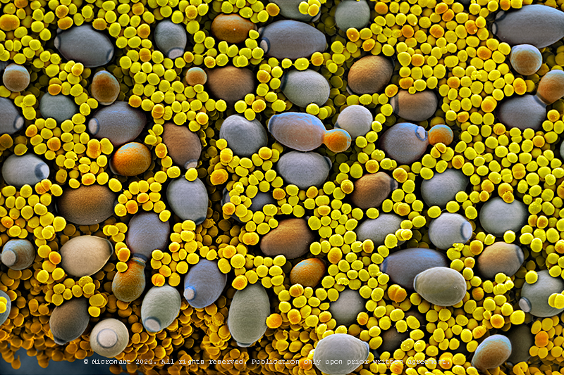
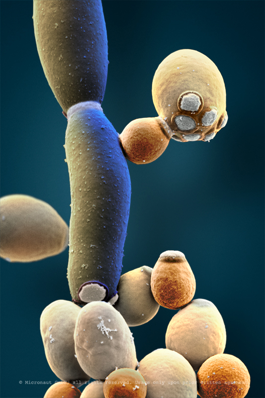
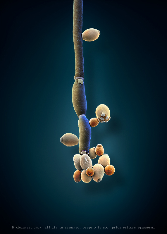
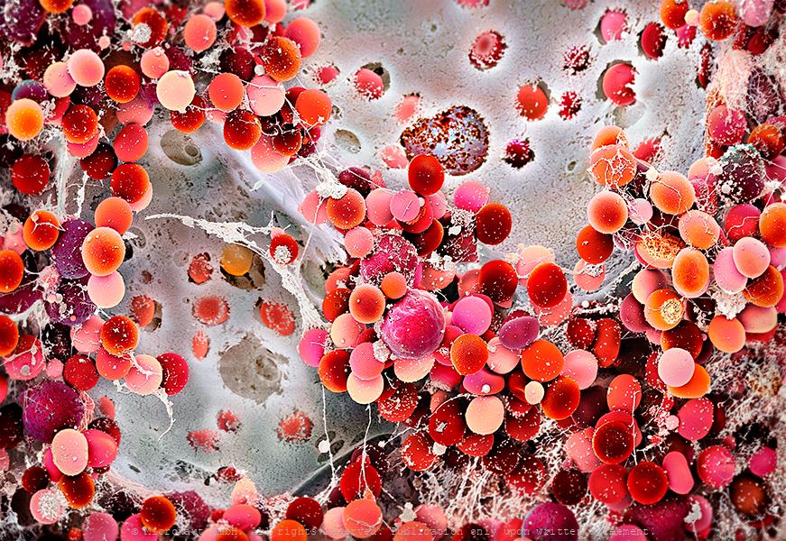
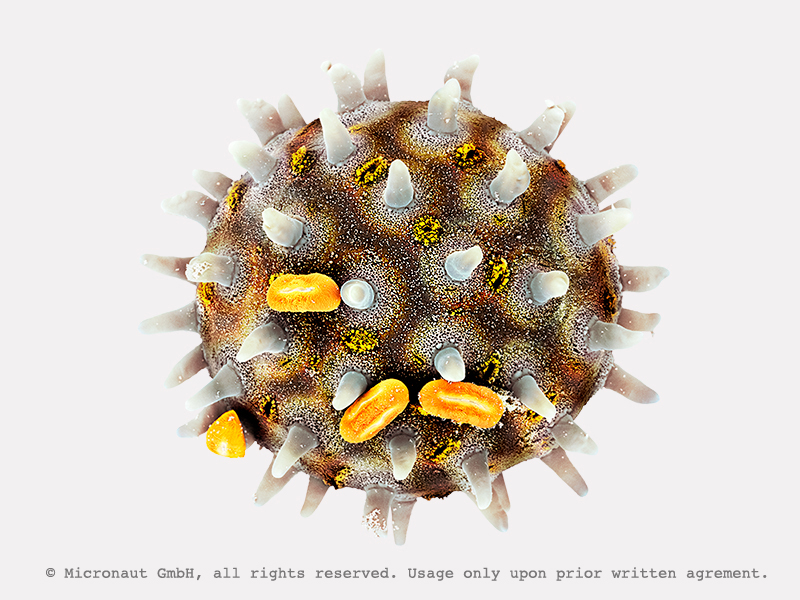
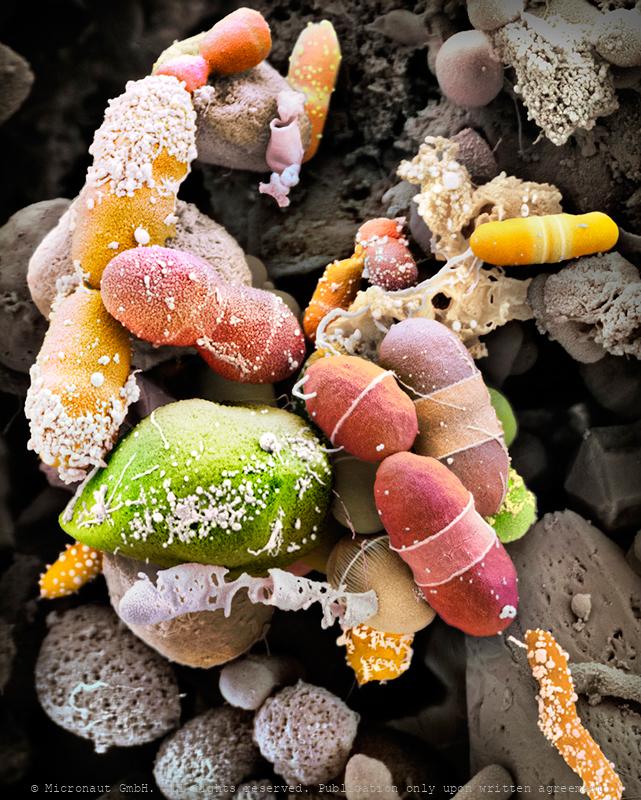
Blastoconidia of Candida albicans
Candida albicans (laboratory culture), hand-colored SEM scan by Martin Oeggerli (Micronaut). Candida albicans is the most common member of the human gut flora and it can also survive outside the human body. In healthy human adults, it is a commensal microbe of the gastrointestinal tract and mouth in 40–60%. It is a multi-morphologic creature with highly different structures: under certain environmental factors blastoconidia are produced. They are very small cells (the yellow cells) in this picture, produced asexually. Blastoconidia are able to divide and further spread within very short time. Despite it is a very common commensal species, C. albicans can also become pathogenic in immunocompromised individuals, causing the human infection candidiasis, which results from an overgrowth of the fungus and can often be observed in e.g. in HIV-infected patients. Candidiasis is causing 2800-11200 deaths in the US yearly. Furthermore, C. albicans is the most common fungal species isolated from biofilms. It is also used as a model organism to study fungal pathogens. According to new studies, C. albicans can cross the blood–brain barrier in mice. Interestingly, C. albicans can grow as both, yeast (opaque and GUT form) and filamentous cells (pseudohyphal form): due to limited mobility, fungi have evolved mechanisms to adapt to sudden chemical and/or physical variation in their environment and scientists are using C. albicans to study eukaryotic responses to environmental changes by switching morphologies from yeast to hyphae growth.
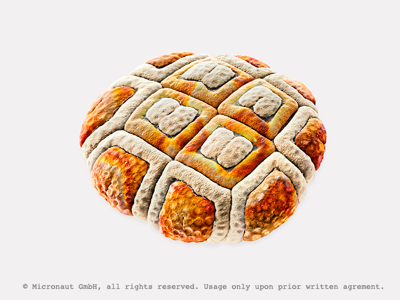
Face/Off - The interchangeable fungi (Candida albicans)
Candida albicans (laboratory culture), hand-colored SEM scan by Martin Oeggerli (Micronaut). Candida albicans is the most common member of the human gut flora and it can also survive outside the human body. In healthy human adults, it is a commensal microbe of the gastrointestinal tract and mouth in 40–60%. However, C. albicans can also become pathogenic in immunocompromised individuals, causing the human infection candidiasis, which results from an overgrowth of the fungus and can often be observed in e.g. in HIV-infected patients. Candidiasis is causing 2800-11200 deaths in the US yearly. Furthermore, C. albicans is the most common fungal species isolated from biofilms. It is also used as a model organism to study fungal pathogens. According to new studies, C. albicans can cross the blood–brain barrier in mice. Interestingly, C. albicans can grow as both, yeast (opaque and GUT form) and filamentous cells (pseudohyphal form): due to limited mobility, fungi have evolved mechanisms to adapt to sudden chemical and/or physical variation in their environment and scientists are using C. albicans to study eukaryotic responses to environmental changes by switching morphologies from yeast to hyphae growth.
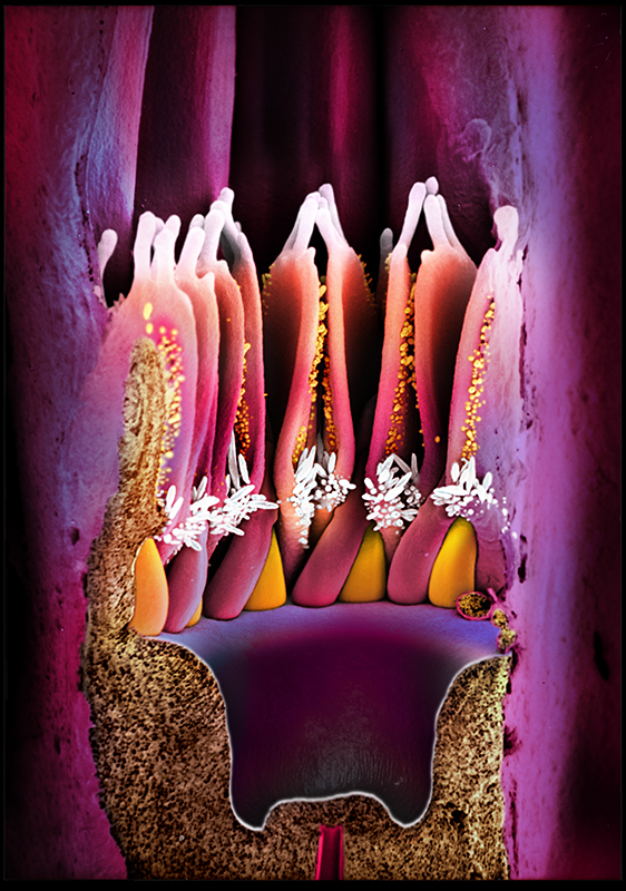
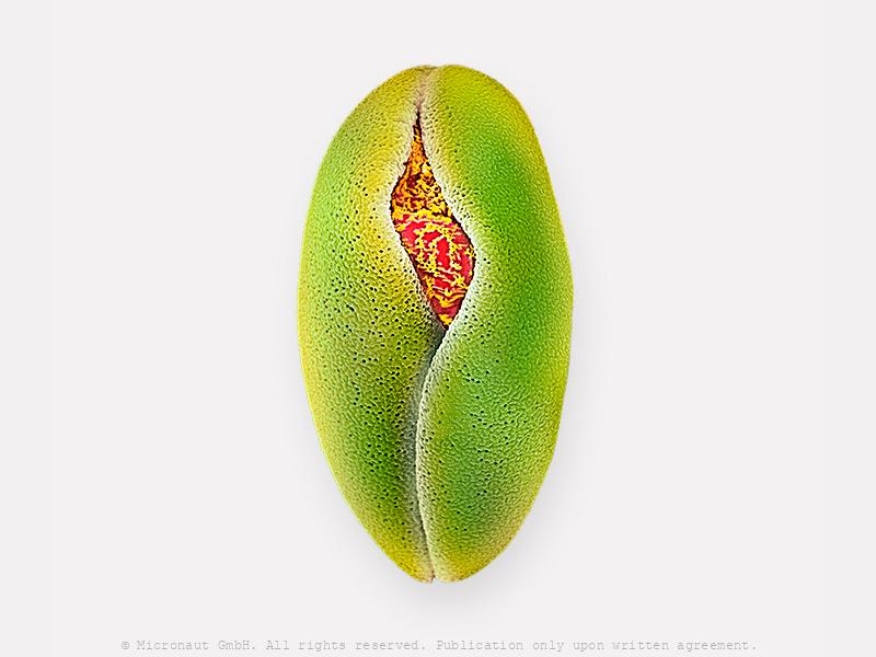
Floral Elements (Bruguiera gymnorhiza) - Nr.1
The species is Bruguiera gymnorhiza produces red pendulous flowers which attract birds (hence, bird pollination). This picture shows a longitudinal section through a young flower: at the very bottom you can see the "basal wing" of a young petal that guides a young stamen inside the petal. Next the curved stamen will elongate upwards. Reference page: http://iriomote.image.coocan.jp/research/mangrove/Bruguiera/BruguieraEnglish.html
Solar Powered Future (C. staurestrum)
Image shows C. Staurestrum algae from Prof. Ben Hankamer's multidisciplinary approach to use light-driven microalgae chemistry to convert solar energy into sustainable products and ecosystem services. This breakthrough “third way” of harnessing the sun's power goes beyond electricity and heat, transforming sunlight directly into renewable fuels, food and clean water. The team’s work addresses urgent global challenges including CO₂ reduction, food security and water purification, while leveraging Australia’s natural strengths — sunlight, space and scientific talent. With solar energy capable of powering the world in just two hours, solar biotechnologies offer a scalable path to new green industries, jobs, and exports. Driven by purpose and innovation, Professor Hankamer’s research positions Australia as a global leader in sustainable solar solutions, demonstrating how science can turn energy abundance into environmental and economic resilience.
Porcine retina (Sus domesticus)
Porcine Retina (Sus domesticus) With far more than 100 million nerve cells, the retina is our first stage of the visual system and window to the outside world. The picture highlights the neuronal layers of a porcine retina. Seungkuk Ahn and his colleagues have electromagnetically guided viruses to specifically transduce each of the retina layers. The process inside the retina includes detection of light impulses (photons) by different light receptors, in general called rods (120 million cells) and cones (6 million cells), and the fast and continuous translation, filtration and post-procession into electrical signals (or nerve impulses). These signals are then passed through the optical nerve’s 1.5 million fibres to the visual centre of the brain and reinterpreted into a cohesive image.The flexibility and economy with which retinal cells work together remains beyond our powers of imagination: the eye reports numerous signals to the brain at once, including separate detection of light (light on) and dark (light off), general patterns and finest details, movements and different hues (including: red, green and blue). This literally makes the eye a camera with 10 to 15 different films and despite it appears so naturally to us, the perception of an image is the result of a most complex interaction process involving millions of cells and the electrical signals they produce and pass on to the brain.
Face/Off - The interchangeable fungi (Candida albicans)
Candida albicans (laboratory culture), hand-colored SEM scan by Martin Oeggerli (Micronaut). Candida albicans is the most common member of the human gut flora and it can also survive outside the human body. In healthy human adults, it is a commensal microbe of the gastrointestinal tract and mouth in 40–60%. However, C. albicans can also become pathogenic in immunocompromised individuals, causing the human infection candidiasis, which results from an overgrowth of the fungus and can often be observed in e.g. in HIV-infected patients. Candidiasis is causing 2800-11200 deaths in the US yearly. Furthermore, C. albicans is the most common fungal species isolated from biofilms. It is also used as a model organism to study fungal pathogens. According to new studies, C. albicans can cross the blood–brain barrier in mice. Interestingly, C. albicans can grow as both, yeast (opaque and GUT form) and filamentous cells (pseudohyphal form): due to limited mobility, fungi have evolved mechanisms to adapt to sudden chemical and/or physical variation in their environment and scientists are using C. albicans to study eukaryotic responses to environmental changes by switching morphologies from yeast to hyphae growth.
Der Zauberpilz - The Psycodelic Mushroom (Claviceps purpurea)
Lysergic acid diethylamide is a synthetic lysergamide that can be obtained as a derivative of naturally occurring ergot alkaloids. LSD is one of the most powerful hallucinogens known and belongs to a subgroup of psychedelics that affect the body's serotonin system. In mid-April 1943, the Swiss chemist Albert Hofmann accidentally discovered the psychoactive substance LSD. The drug has since been banned, but in the right dosage and with therapeutic support, LSD can also be used as a helpful medication against depression. Thinking, feeling and perception are massively influenced by the psychoactive substance, the experience of space and time changes, sensory illusions, colors, image distortions or delusions may occur. -- Lysergsäurediethylamid ist ein synthetisches Lysergamid, das als Derivat natürlich vorkommender Mutterkornalkaloiden erhalten werden kann. LSD ist eines der stärksten bekannten Halluzinogene und gehört zu deren Teilgruppe der Psychedelika, welche auf das Serotonin-System des Körpers wirken. Mitte April 1943 entdeckte der Schweizer Chemiker Albert Hofmann per Zufall die psychoaktive Substanz LSD. Zwischenzeitlich wurde die Droge verboten, doch in richtiger Dosierung und in therapeutischer Begleitung kann LSD auch als hilfreiches Medikament gegen Depressionen eingesetzt werden. Denken, Fühlen und die Wahrnehmung werden durch die psychoaktive Substanz massiv beeinflusst, das Erleben von Raum und Zeit verändert sich, es kann zu Sinnestäuschungen, bunten Farben, Bildverzerrungen und Wahnvorstellungen kommen.
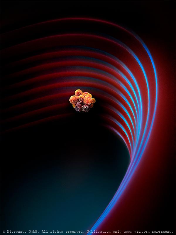
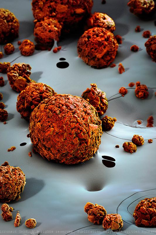
Cancer spreads during sleep (Circulating Tumor Cell Cluster)
Circulating Tumor Cell Cluster (CTC cluster; metastasis) on a microfluidic device
Satellite Cell (Mus musculus) - The Stem Cell that Regenerates t
2021 marks the 60th anniversary of the discovery of the "satellite cell" by Alexander Mauro by using the electron microscope and it seems an opportune moment for two researchers from the University Basel to team up with Micronaut and present a state of the art picture of a muscular stem cell. To produce this picture, mouse muscles were digested and cultured by Sedat Dilbaz and Gesa Santos in the lab of Prof. Christoph Handschin, at the Biozentrum, University Basel. Once satellite cells got activated, the sample was carefully fixed and a scanning electron microscope (SEM) was used to capture black-and-white SEM scans. After this, photorealistic colors were added in the digital darkroom by the artist. The basic unit of skeletal muscle is the myofiber: a syncytial cell packed with myofibrils, containing the sarcomeres that generate force by contraction. In vertebrates, each myofibre is controlled by many (usually hundreds) of myonuclei, which in mammals are considered post mitotic. During growth, new myonuclei are supplied by muscle satellite cells: resident stem cells located on the surface of a myofibre. In mature muscles, satellite cells become quiescent ("dormant"), but remain able to provide myoblasts for muscle hypertrophy and repair if needed. That muscle is capable of regeneration was first shown in the 1860s, but almost a century elapsed before the satellite cell was discovered. Electron microscopic studies by Alexander Mauro identified the satellite cells wedged between the plasma membrane of the muscle fibre and basement membrane and revealed that they almost completely lack cytoplasm. While nowadays, the muscle satellite cell is widely accepted as the resident stem cell of skeletal muscle, supplying myoblasts for growth, homeostasis and repair, less is known about how Satellite cells remain quiescent, or how they are activated. The understanding of the complex cellular and molecular interactions of satellite cells with their dynamic microenvironment rem
Blood- and bone marrow stem cells (Homo sapiens) - Knochenmarkas
A stem cell or bone marrow transplant replaces damaged blood cells with healthy ones. It can be used to treat diseases such as leukaemia and lymphoma, which affect the blood cells of the patient. After removing damaged blood cells, healthy cells can be derived from blood- or bone-marrow (a spongy tissue found in the centre of some bones) stem cells. Stem cells can turn into different types of blood cells, including: - red blood cells – which carry oxygen around the body - white blood cells – which help fight infection - platelets – which help stop bleeding. By Martin Oeggerli, supported by SRK, Andi Buser and Andi Holbro, and Pathology Univ. Hosp. Basel, and Bio-EM Lab, Univ. Basel.
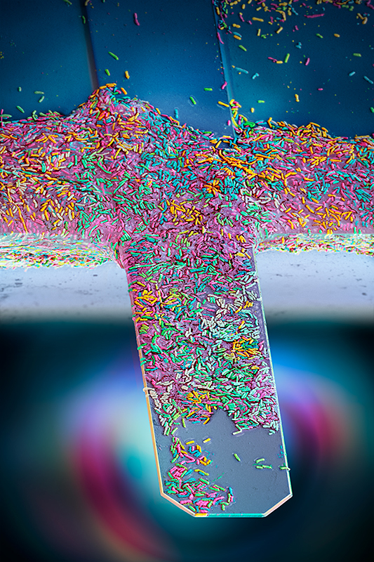
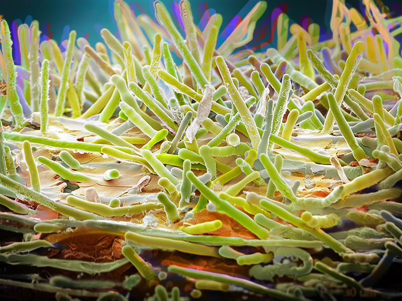
Nanomotion sensor - detecting the vibration of living bacteria i
Antibiotic Resistance Chip Loaded with Bacteria. Hand-colored SEM scan by Martin Oeggerli / Micronaut, 2021. Antibiotic resistance is currently one of biggest threats to global health. By offering a diagnostic device, Resistell proposes an alternative to culture based antibiogram, the current gold standard in antibiotic susceptibility testing. The rapid AST method is based on the detection of movement caused by living bacterial cells. Because the test is growth independent rapid AST, we reduce the time taken to get a result from days to minutes. Resistell provides information on which antibiotic should be used to treat the patient, and the concentration at which it should be administered. This rapid AST designed and developed by Resistell saves lives by finding the right antibiotic on time and reduces spreading of antibiotic resistance!
Bone Structure (Mus musculus)
This portion of the Petrous bone (Mus musculus) is protecting the inner ear structure. Though it is one of the hardest, densest bones in the body. Petrous comes from the Latin word petrosus, meaning “stone-like, hard”.
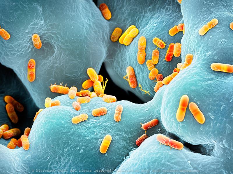
The human gut flora - bacteria (Enterobacter cloacae)
Enterobacter cloacae bacteria, coloured scanning electron micrograph (SEM). Enterobacter cloacae is a clinically significant Gram-negative, facultatively-anaerobic, rod-shaped (bacillus) bacterium. Members of the genus Enterobacter exist in water, soil and the intestinal and urinary tracts of humans. In hospital patients with lowered immunity E. cloacae may be the causative agent of urinary tract and respiratory tract infections. In addition, it may also infect wounds on the skin, and quite often shows multiple resistancy to antibiotics.
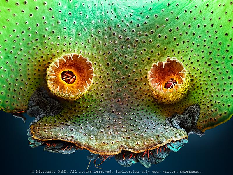
Hearing Sensors (Mus musculus)
Hearing Sensors This image shows the sensory epithelium of the inner ear (Organ of Corti), with the apical protruding hair bundle of hair cells. Scanning-electron-microscopic (SEM) image of a 22-week-old wild-type C57BL/6J mouse, carefully colored to harmonize with the natural shadows of the image and to highlight the invisibly small structures that are responsible for hearing in mammals. The organ of Corti was called after the Italian scientist (Alfonso Corti), who first described it. Its three rows of highly ordered hair cells, located inside the cochlea, are unique among other organs of the body and generate nerve impulses in response to sound vibrations. By Maurizio Cortada and Martin Oeggerli. Supported by Pathology, Univ. Hosp. Basel, and Bio-EM Lab, Univ. Basel.
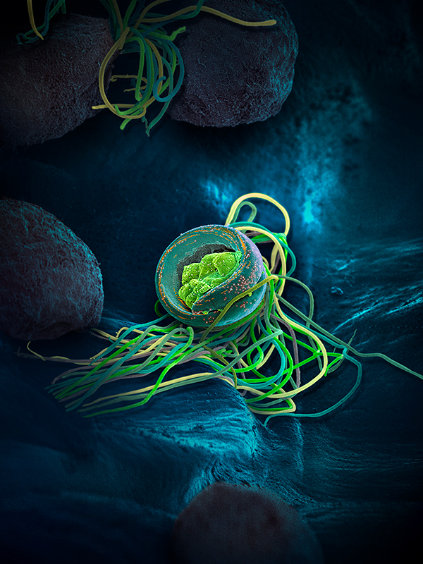
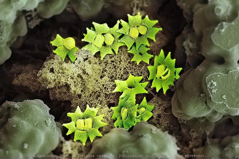
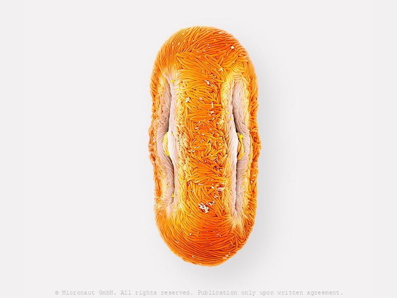
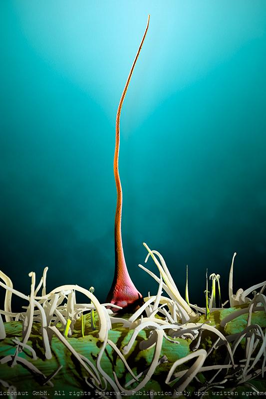
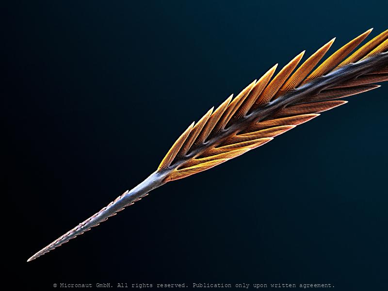
A Sperm is Born - Sword Fern (Polystichum munitum)
Many-flagellated sperm of the swordfern, Polystichum munitum. The nucleus (in blue with orange flecks) is in a condensed spiral with the remaining cellular organelles at the tip of the sperm (in green). Various long flagellae drive the sperm through the water to find the egg. While flowering plants have developped pollen grains to protect their male gametes on their epic journey to fertilize the egg cell, ferns and mosses produce motile gametes (similar to animals) which is unique in the kingdom of plants. The immense majority of animals and plants reproduce sexually. In sexual reproduction, haploid cells called gametes combine during fertilization to result into a diploid zygote that starts to divide and develop into an embryo. Sperm and egg cells are invisibly small and only present at the initial phase of a creatures existence. Thanks to experts and high-tech equippment, it is possible to shed light on this critical moment at the "start of life", which maintains genetic variation and allows organisms to evolve and adapt to their environment. Scanning-electron-microscopy (SEM) image captured in collaboration with Patrick von Aderkas, Elaine Humphrey and Brent Gowen. Hand-colored artwork by Martin Oeggerli (Micronaut).
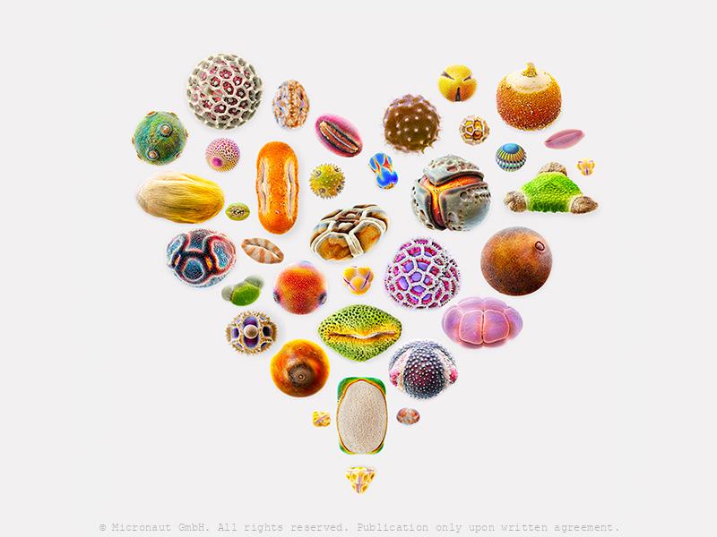
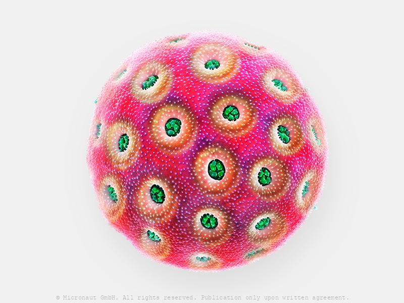
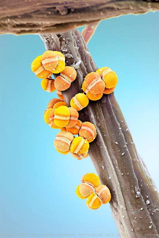
Valentine's Day - Assortment of pollen grains
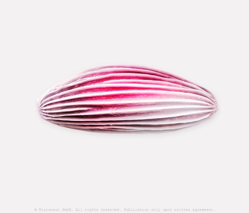
Water Cabbage Pollen (Pistia sp.)
Water cabbage (Pistia sp.) is an invasive plant and was first described from Nile close to lake Victoria. Despite a tiny flower, the pollen is a medium sized elliptic monad covered with equatorial crests, which are rarely found features in the pollen universe.
David versus Goliath (Pollen grains of Hibiscus sp., and Anethum
Hibiscus (Hibiscus sp.) pollen grain with four Dill (Anethum graveolens) pollen sticking to its surface. Hibiscus blossoms are brightly colored and open wide, attracting pollinators from bees to hummingbirds to reach in for a deep drink of nectar. As they do, they inevitably make contact with the flower's prominent stamen and pistil, aiding in the plant's pollination and subsequent fertilization. The pollen of Hibiscus is one of the largest that you can find in the pollen universe and it cannot be transported by the wind. Instead, it relies on elongated spikes on the surface, which are considered a characteristic adaptation to insect (& bird) pollination, to facilitate its attachment to hairs and feathers. Since Hibiscus pollen are far too heavy to be dispersed by the wind, it cannot be considered an allergenic threat. However, if you are extra sensitive you may want to avoid Hibiscus tea as it can contain irritants.
©-Micronaut-Pollen-Acacia-001028
Tape Amaranth pollen grain (Amaranthus sp.)
Amaranth (Amaranthus sp.) pollen grain. Pollen is a fine to coarse powder consisting of microgametophytes (=pollen grains), which produce the male gametes (sperm cells) of seed plants. Insect pollinated plants produce relatively low numbers of pollen grains compared to wind pollinated species. Each plant species has pollen grains that are unique due to the meticulous processes of evolution. While some have only one aperture (pre-formed opening to let the pollen tube exit), others may have three, six, or even double and triple the number. To have many pre-formed openings is assumed to be a more modern development: it allows the delicate pollen tube to emerge and dive right into the pistil tissue, without having to grow over an extensive stretch while being exposed to UV radiation, heat and other stress factors that could lead to damage of the tube and male gametes. Oeggerli slowly breathes life into his works by manually highlighting different structures with colors, layer upon layer on his laptop. The process allows him to set focus on the mysterious diversity and hidden morphological structures of pollen grains. by Martin Oeggerli and Louisa Howard, Microscopy Facility, Dartmouth College.
Dill pollen (Anethum graveolens)
Dill (Anethum graveolens) pollen grain. It's an elongated, relatively small to medium size pollen grain featuring three apertures (i.e. pre-formed openings for exit of the pollen tube). Pollen grain by Martin Oeggerli and Louisa Howard, Microscopy Facility, Dartmouth College.
Snowdrop pollen (Galanthus nievalis)
Common snowdrop (Galanthus nievalis) is a typically wind pollinated plant. In early spring, the plant grow from bulbs –sometimes directly through snow- to open just a single flower. Pollen are oval shaped with a smooth surface.
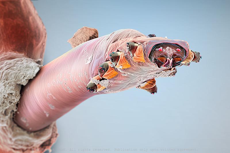
The Lodger - Face Mite (Demodex)
Demodex is a type of mite that lives in human hair follicles, usually on your face. Almost everyone has these mites, but they usually don’t cause problems, except if Demodex can multiply too quickly which is the case in people who are immunocompromised or have other skin conditions. In such a case, face mites can cause an itchy, irritating condition called demodicosis. The parasite is well adapted to its environment through thousands of years of co-evolution with Homo sapiens and extremely tiny (0.15 - 0.4 millimeters). It would take several of them to cover a pin head. Under a microscope, the mite looks slightly transparent and is often covered with scales. It has an elongated body with two segments. The first segment has eight legs and a mouth. When you sleep, the mites come out of your skin’s pores, mate, then go back into your skin to lay eggs. Demodex mites can spread from human to human. Babies aren’t born with mites, but they get mites from the people they live with. Two main species of Demodex live on humans: - Demodex folliculorum: usually lives in smaller hair follicles, especially your eyelashes. They eat skin cells. - Demodex brevis: usually lives near the oil glands in hair follicles. They eat sebum, a greasy substance made by oil glands. This is a photorealistically colored scanning electron micrograph by Martin Oeggerli. The artist also uses colors to make visible specific structures.
The most primitive plant on earth - Liverwort (Marchantia polymo
LIVERWORTS are untypical non-vascular plants. They look very much like flattened moss, and measure only between 2-20mm. Due to the small size, liverworts are often overlooked, despite there are about 9.000 species. As a gardener you may wondered how they sometimes become so widely established in your greenhouse - here's why: a) The flat green leaf-like body of the liverwort spreads horizontally across the media surface by dividing into two branches at apical notches. Each apical notch contains a growing-point (called 'meristem'), that can growth laterally if there is media to grow on. Fragments that are dislodged or torn away from the mother plant then have the potential to become independent new plants. b) In addition, the upper surface of the liverwort features 3-4mm cup-like structures called gemma cups. They contain over 100 tiny, disk-shaped gemmae, which represent clones of the mother plant. When water droplets hit the cups, the tiny gemmae are propelled out, sometimes up to 15 cm in the air, and each gemmae can eventually develop into a new plant if the conditions of living fit. c) Finally, liverworts which are either male or female plants, can also spread through sexual reproduction. In the presence of water, sperm may swim from the male plant up on a nearby female structure and fertilize the ovum which develops into a spore that will be eventually be spread by air currents. Female plants produce spores in the thousands and they are very small (less than 3.5µm in diameter). Starting with Greek philosophers such as Aristotle and Theophrastus, liverworts have been mentioned in the herbal literature (in many cases as 'lichen'). In the Middle Ages in Europa the species Marchantia polymorpha was thought to be useful for treating diseases of the liver, based on the plants morphologically which resembles the human organ. Nowadays, liverworts are an interesting topic in modern research, because they stand at the very beginning of the plant tree of life and com
Stinging Vegetable Velcro (Eucnide bartonionides)
The Stingbush (Eudnide bartonionides) is a plant is a shrub which is native to desert areas in California, Arizona, Utah and Baja California. The leafs are equipped with short non-stinging ('velcro') hairs and special elongated stinging hairs (center). Stinging hairs (trichomes) produce a painful stinging sensation by injecting a chemical mixture when touched by humans or other animals. They act like hypodermic needles: after the tip breaks off, a chemical mixture composed of different chemicals that are injected and cause pain or paresthesia. Shorter 'velcro' hairs hold on your skin if you are touching the plant leaf, to increase damage and pain caused through elongated stinging hairs.
Synthetic Diamonds
Synthetic Diamonds A natural diamond is made from carbon and is the hardest natural known substance on earth. Natural diamonds are created over a period of one to three billion years, at least 85 miles below the earth’s mantle under natural conditions of very high pressure and high temperature. Once a diamond has been created in these underground conditions, it travels via molten rock to the earth’s surface, where it is mined, refined, and turned into beautiful jewelry or used for industrial purposes. Natural diamonds are slightly more durable, ranking 10 out of 10 on the Mohs Hardness scale, whereas synthetic (lab-) diamonds rank 9.25.
Capsugel boulders
Capsugel - Lonza - Pharmaceutical
human Microbiome of a KISS
The Human Microbiome of a Kiss. Artwork created by Martin Oeggerli (Micronaut) and dedicated to his 8-years old son Nelson. This image was originally produced for the National Geographic feature article 'How trillions of microbes affect every stage of our life-from birth to old age'. Soon after its public release, our daily life became heavily overshadowed by the world's coronavirus (COVID-19) pandemic - thereby instantly confirming the articles visionary title. Meanwhile (at end of April 2020), the crisis has not only severely limited the artists ability to work, but also prohibits him from officially visiting his son who is living in Switzerland, just a stone's throw away from the rest of the father's German-based patch-work family. This picture is based on scanning-electron-microscopy technology, and all colors are manually added by the artist in post-production, to visualize the immense diversity of the microbial community transmitted through a kiss. Whenever we kiss someone on the lips or cheeks invisibly small microbes are exchanged from one person to the other one! The variety of microbes that are part of our microbiome is mind-blowing, and the composition of the invisibly small communities can be the cause of many factors, including the human genetic makeup, diet, age, surroundings, and sexual behavior. Among humans, approximately 90% of cultures have some type of kissing. Usually it is platonic, such as a parent kissing a child. However, in 77 of 168 (46%) of all cultures, it can go as far as intimate (French-) kissing. Recently, a scientific study has revealed that on average 80 million bacteria are transferred to the partner during a kiss of 10 s. Most partners share a more similar oral microbiome compared to unrelated individuals and/or e.g. their kids. But some of the collective bacteria among partners are only transiently present, while others have found a true niche and survive permanently, allowing long-term colonization.With regard to kissing, th
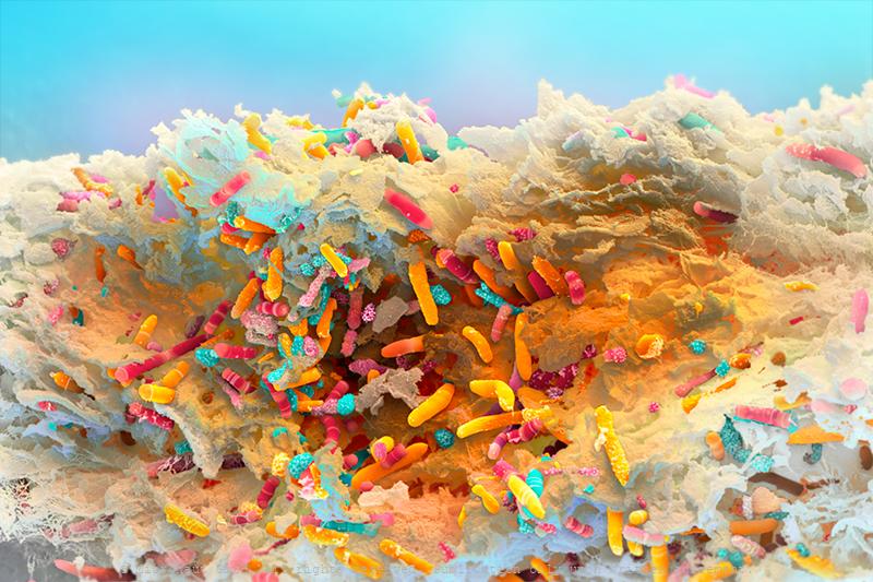
The Gut Microbiota Miracle - Microbiome of a Newborn (H. sapiens
The microbiome of a baby, 1 month after birth. Hand colored scanning electron micrograph by Martin Oeggerli (Micronaut). The Gut Microbiota Miracle: this picture shows the microbiome of a newborn one month after birth. The importance of our intestinal bacteria is already well known – but much less is known about its development and diversity from a newborn to the adult. It has long been assumed that human breast milk is sterile. But scientists have discovered complex and dynamic bacterial ecosystems in human breast milk which are distinct from other microbioal communities and may also colonize and widen the diversity of the infant gut probiotic bacterial community. According to the study, the cocktail of beneficial bacteria passed from mother to infant through breast milk changes significantly over time and could act like a daily booster for infant immunity and metabolism, with important implications for infant development and health. Bacteria found in breast milk include Actinobacteria, Bacillariaphyta, Bacteroidetes, Cyanobacteria, Deinococcus-thermus, Firmicutes, Fusobacteria, Patescibacteria, Plantae, and Proteobacteria.
The Human Microbiome, Nr.2
Excrements revealing the bacterial flore of the human intestinal tract. If it comes to digestion, we are not alone! Among many more bacterial forms streptococci, staphylococci, enterococci, enterobacteria, mycobacteria, spirochetes, mycoplasma, corynebacteria, clostridia, or lactobacilli are living in our gastro-intestinal tract and usually help us to digest food, aid nutrient absorption, reduce dehydration or assist in the production of key vitamins. In adults, the bacterial flore makes up about five percent of the total body weight (similarlly to the brain). Altogether, more than 30'000 different species of bacteria have been classified in our intestinal tract, so far! However, it’s a fragile ecosystem. The bacterial flore is sensitive to stress, unhealthy food, alcohol or illness. Under unfavorable circumstances it directly affects our health and e.g. causes overweight, changes our mood or makes us prone to various diseases. The small intestine normally contains relatively low numbers of bacteria compared to the large intestine. If abnormally large numbers of bacteria grow in the small intestine, they may use many of the nutrients that a person would normally absorb for their growth. Too much growth and breakdown of nutrients can damage the cells of the small intestinal wall, leading to Crohn’s disease, diabetes, scleroderma and severely influence the progression of AIDS or immunoglobulin deficiency. This hand-colored scanning electron micrograph reveals the large number and variability of bacteria that are present in human excrements. Additionally, a plant fiber has passed the intestinal tract almost unharmed and spans across the frame (center). On the right hand side (half-cut) a large and spherical shaped structure turns out to be the cyste of a Giardia parasite (brown) - the actual reason why the person went to see a doctor... But apart from the unusual parasitic supplement the bacterial fauna looks fairly well-sorted.
Anthrax (Bacillus anthracis)
Anthrax bacteria (Bacillus anthracis) is a Gram-positive endospore-forming rod more commonly called “anthrax”. In 2001 the organism was used as part of a terrorist attack on the USA, where a highly virulent strain of anthrax was mailed to senators killing 5 people. This has heightened the concern around anthrax as a biological weapon (Schwartz, 2009). B. anthracis is commonly found in soil where it can lie dormant, potentially for hundreds of years. Herbivores often disturb soil when grazing and can ingest or inhale the B. anthracis spores. The bacteria can be transmitted readily from close contact with animals, soil or through ingestion of contaminated meat (from an infected animal) (Schwartz, 2009). Occupations at high risk of infection include veterinary surgeons, livestock farmers, butchers and persons who handle hides or wool. Treatment and antibiotic resistance: Contrary to popular belief, antibiotics are very effective in controlling B. anthracis infections if used early enough. Antibiotics such as ciprofloxacin, doxycycline, erythromycin, vancomycin and penicillin are all useful for treatment of B. anthracis infections (Moayeri and Leppla, 2004). There is also a monoclonal antibody (Raxibacumab) that is currently showing promise in clinical trials , which can be used in the prophylaxic treatment of B. anthracis infection. B. anthracis has shown some resistance to antibiotics in laboratory testing and possesses antimicrobial enzymes such as b-lactamases (Bryskier, 2002). Prevention and control: Preventative measures include immunisation of humans and animals and post exposure antibiotic prophylaxis (Bryskier, 2002). The currently available vaccines include AVA (anthrax vaccine adsorbed), which is licensed in the USA and AVP (anthrax vaccine precipitated), which is licensed in the UK; these are used to create immunity to B. anthracis pre-exposure (Bryskier, 2002). Where people or animals have died from B. anthracis, great care must be taken to handle
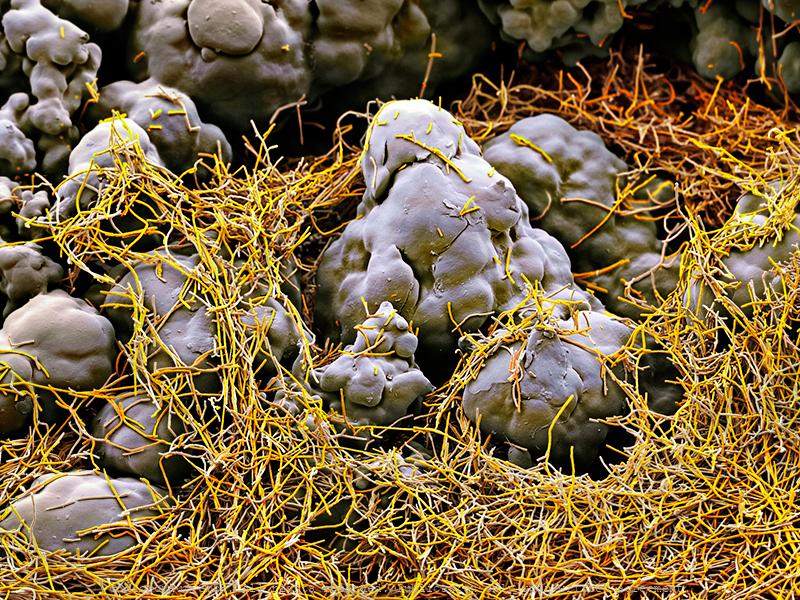
A Fuzzy Jacket for Cheese!
Artwork created by Martin Oeggerli (Micronaut). The picture is based on scanning-electron-microscopy technology, and all colors are manually added by the artist in post-production, to visualize characteristic microstructures of the microbiotic fungus Geotrichum candidum. The fungus G. candidum is a member of the human microbiome, notably associated with skin, sputum and feces where it occurs in 25–30% of specimens. It is also common in soil and has been isolated from soil probes collected across all continents. Furthermore, this yeast is the causative agent of geotrichosis. Pulmonary involvement is the most frequently reported form of the disease, but bronchial, oral, vaginal, cutaneous and alimentary infections have also been reported. G. candidum is also widely used in the production of certain dairy products including rind cheeses such as Camembert, Saint-Nectaire, Reblochon and other wrinkly cheeses that you may find at your local cheese shop. In a Nordic yogurt-like product known as viili, the yeast can be found as well, being responsible for the product's velvety texture. Furthermore, G. candidum has been reported as an organism that is able to break down plastic. Using in-depth genomic sequencing, French scientists recently unlocked the evolutionary history of this important cheese microbe and revealed a fungus with an identity crisis: all true yeasts are descendants of molds. They evolved a single-celled lifestyle from their moldy multi-cellular ancestors. Throughout this evolutionary process, they let go of habits such as growth using the filaments that create the fuzzy appearance of most molds. For a long time, Geotrichum candidum (hereafter G. candidum) has been a mycological mystery to both cheese makers and scientists. It has a fuzzy appearance when it grows on the surface of cheeses or on Petri dishes in the lab, but when you look under the microscope, it also has the appearance of tiny single cells that look like a yeast. And when scientists st
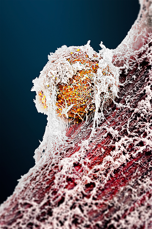
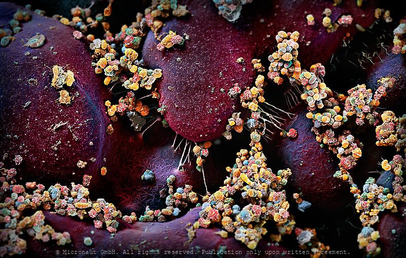
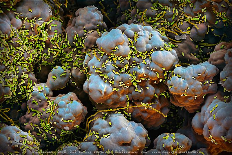
Stomach cells infection - Helicobacter pylori
In vitro cultured stomach cancer cells (red/brown), infected with Helicobacter pylori (yellow). Magenepithelzellen wurden in vitro gezüchtet und experimentell infiziert mit Helicobacter pylori; die Bakterien sind in gelb zu sehen, die Wirtszellen sind rot-braun. In vitro cultured stomach cancer cells (brown), infected with Helicobacter pylori (yellow). Helicobacter pylori, a common bacterium that lives in the stomach lining, increases the risk of stomach cancer and peptic ulcers. But over time H. pylori can reduce stomach acid and acid reflux, which may help fend off esophageal cancer. The microbe also appears to help protect us from allergies and asthma. Some scientists suspect that the dramatic increase in those conditions in the industrialized world could be related to the decreasing frequency of H. pylori in our stomachs, which is partly due to high doses of antibiotics in childhood.
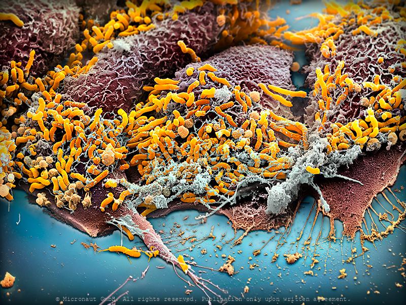
Stinky Foot Bacteria
What causes smelly feet? Sweat is released throughout the day to keep the skin moist and supple. Sweat itself is odorless, but creates a beneficial environment for certain bacteria species to grow and produce bad-smelling substances. The same species are naturally present on our skin as part of the human flora, but in rather low numbers. Some shoes and socks can inhibit evaporation and thus increase the amount of sweat you produce, thereby providing the perfect environment for 'stinky-feet-bacteria' to thrive. Since our feet have more sweat glands than on any other part of the body they typically start to smell bad first.
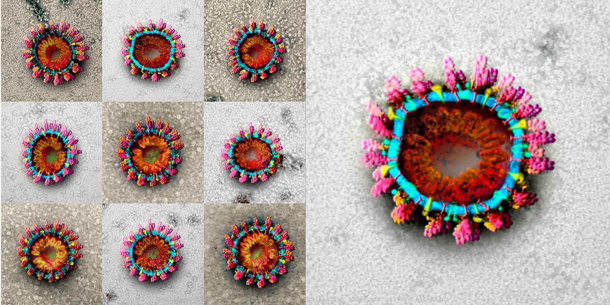
Coronavirus Structures - CoV2
Coronavirus structures. 9 original TEM image scans, hand-colored by Martin Oeggerli.
Pixie dust with a rub in it (Brachypelma smithii)
Ever heard of urticating hairs? Tarantulas have the ability to bite, but urticating hairs are the invisible enemy. Many species of New World tarantulas (i.e. North and South American), can rapidly kick-up urticating hairs from their abdomens using their fourth legs in a rapid motion. The tiny hairs gently float up into the air like pixie dust. But pixie dust it is not. Avoid breathing the bristles or touching your eyes if you have any bristles on your fingers, as it can cause irritation. The urticating bristles can actually be fatal to rodents who inhale them. The urticating hairs themselves look like minuscule floating lint or dust to the naked eye. Under a scanning-electron-microscope they look like barbed spears, and there are six types. If allowed to land on your skin, they can sometimes cause redness and irritation, although I’ve personally only experienced this with Theraphosa tarantulas. It’s more annoying than anything, but a defense mechanism to avoid if possible. Some species have extremely mild urticating hairs, imperceptible to humans. Others, like the Goliath bird-eating tarantula (Theraphosa sp.) have urticating hairs that can itch and result in a mild rash. Hydrocortisone usually does the trick to alleviate any itching. Also, a tarantula may deliberately spread urticating hairs at the entrance to its burrow. Another use is to protect an egg sac, as the hairs help annoy would-be predators, such as fly larvae. It’s important to note that many species of tarantula that have urticating hairs available to them do actually never use them when handled. This includes Rose hairs and Redknees.
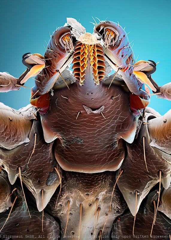
Samurai Armor Protection - The Common Tick (Ixodes ricinus)
Common tick (Ixodes ricinus), hand-colored scanning-electron-micrograph image by science artist Martin Oeggerli. Karuta was a type of armour worn by samurai warriors in Japan. The word karuta comes from the Portuguese word meaning card, (carta) as the small square or rectangular plates that compose the armour resemble traditional Japanese playing cards. In a very similar fashion, the common tick is protected by rigid chitin plates against mechanical damage. Everyone who has ever tried to remove and crush a tick knows very well how tough it is to kill the beast. In addition to the armor-jacket, ticks have also developed other special equipment, e.g. to locate and prepare the right place for the sting (palpi and chelicerae), or to hold on persistently (hypostome and claws), and even resist when the host is moving fast, or is actively trying to remove the parasite by scratching or rubbing. Even more interesting it becomes, if you look at the exquisite details: the barbed stinger (hypostome) features a barrett file structure that is shaped with incomprehensible precision. Each leg ends with a pair of needle-shaped claws, and a fascinating combination of movable splints, adhesion (?) pads, and a mysterious sole which appears to be highly flexible like a towel that can be wrapped around the other structures of the foot. Another high-tech equipment, the Haller's Organ (not visible in this portrait), is located at the front legs and allows the tick to detect chemicals like carbon dioxide, ammonia, and pheromones. It can even sense humidity and infrared light emitted by the warm, blood-filled body a tick wants to find. In-line with the usual allegation of the tick that it can cause infections and spreads diseases (tick-borne encephalitis and/or Lyme disease), this specimen is carrying around a couple of fungi spores and tiny hyphens are sticking to the steel jacket, too. The risk of being bitten by a tick is highest between March and October. Ticks become active a
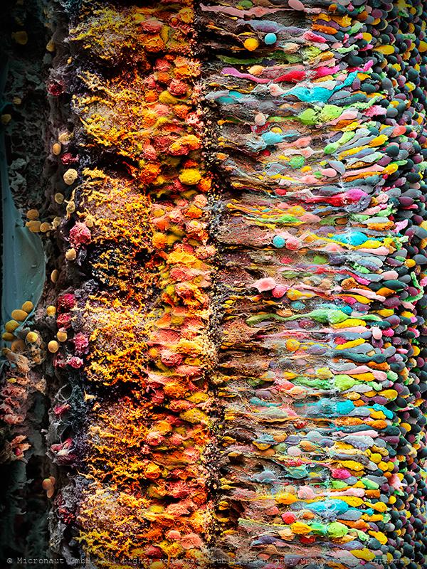
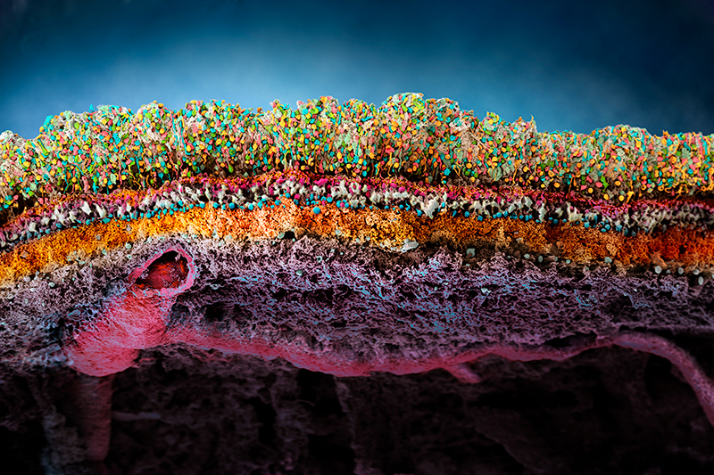
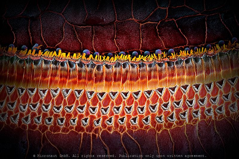
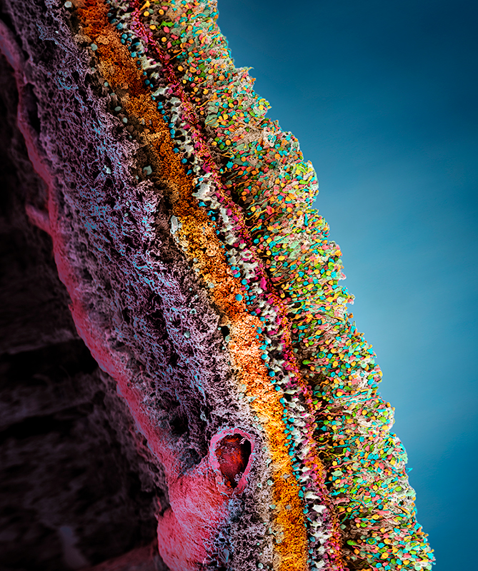
The human Retina - Nr. 3 (2019), vertical
With far more than 100 million nerve cells, the retina is the first stage of our visual system and our window to the outside world. The process includes detection of light impulses (photons) by different light receptors, in general called rods (120 million cells) and cones (6 million cells), and the fast and continuous translation, filtration and post-procession into electrical signals (or nerve impulses). These signals are then passed through the optical nerve’s 1.5 million fibres to the visual centre of the brain and reinterpreted into a cohesive image.The flexibility and economy with which retinal cells work together remains beyond our powers of imagination: the eye reports numerous signals to the brain at once, including separate detection of light (light on) and dark (light off), general patterns and finest details, movements and different hues (including: red, green and blue). This literally makes the eye a camera with 10 to 15 different films and despite it appears so naturally to us, the perception of an image is the result of a most complex interaction process involving millions of cells and the electrical signals they produce and pass on to the brain. What you can see on this image is the (1) rods and cones layer. It is located below the pigment epithelia which has been cut away during the preparation and is not visible on this image (black space on top). Below the rods and cones layer you can find the (2) outer nuclear layer (ONL) which contains the nuclei of the rods and cones. The adjacent (3) outer plexiform layer (OPL) is followed by the (4) inner plexiform layer (IPL), which contains numerous cell types, including horizontal cells, bipolar cells and amacrine cells. At the very bottom you can see the (5) inner nuclear layer (INL), followed by the (6) ganglion cell layer with two (7) blood vessels shown at left and (8) nerve fibers (which form the optical nerve).
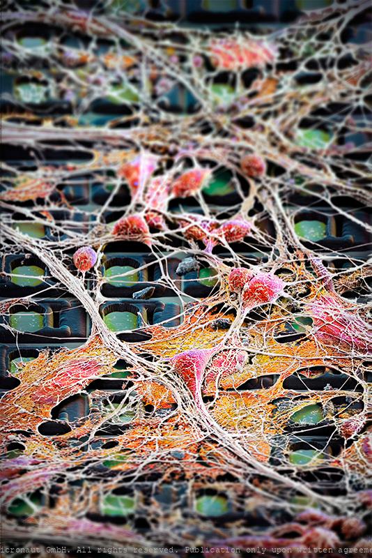
Neuronal Highways - Brain Cells on a Chip
Preparations of electro-active cells, predominantly brain cells, are placed directly atop fully processed microelectronics chips carrying thousands of electrodes. The chips are used to address fundamental questions in neuroscience and medicine. Artwork created by Martin Oeggerli (Micronaut). The picture is based on scanning-electron-microscopy technology, and all colors are manually added in post-production to visualize the research field of Electrophysiology and Neuroscience. We pursue an extracellular, bioelectronic approach to electrophysiology, which relies on the close juxtaposition of electrogenic cells (cells that produce electrical activity) and tissues with state-of-the-art integrated electronic systems. The cell preparations (dissociated cells, tissue slices) are placed directly atop fully processed microelectronics chips carrying thousands of electrodes and featuring CMOS circuitry. The bio-electronic interface consists of noble-metal electrodes. Prospective fields of applications of our technology include, besides fundamental neuroscience research, pharmascreening, the investigation of mechanisms of neurodegenerative diseases, or the development of aural and visual prostheses. Electrophysiology is the study of the electrical properties of biological cells and tissues. It involves measurements of voltage changes or electric currents on a wide variety of scales from single ion channels to whole organs like the heart. In neuroscience, it includes measurements of the electrical activity of neurons and, particularly, action potential activity. Neuroscience is the scientific study of molecular, cellular, developmental, structural, functional, evolutionary, computational, and medical aspects of the nervous system. It is an interdisciplinary science that includes aspects of biology, chemistry, computer science, engineering, mathematics and medicine.
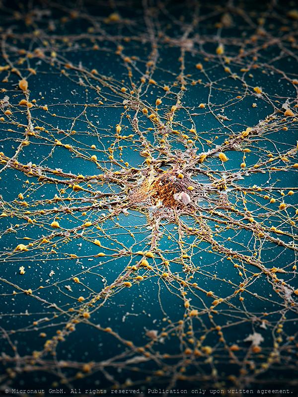
The Neuronal Megacity (Brain cell)
My concept concerning this picture was to compare a nerve cell to an island, or megacity, indicating the complex architecture of the brain. In detail, an infinite number of cellular extensions and microscopical structures unravels (capacity & complexity of the brain), whereas in the overview you can see a confusing tangle of connections that lead towards, or away, or around the center point, like the road network in a megacity. The edges of the picture are deliberately darkened and blurred, indicating there is a lot to be discovered and learned in this area of modern scientific research. Last but not least - the blue “sea” stands for Hawaii / Sophie H, to whom this picture is dedicated. Artwork created by Martin Oeggerli (Micronaut). The picture is based on scanning-electron-microscopy technology, and all colors were manually added to visualize the complex network as formed by neuronal cells in vitro.
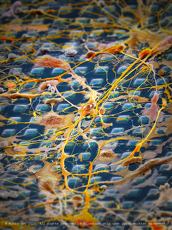
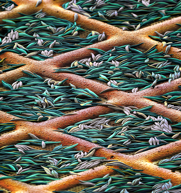
Exploring Cellular Interactions with a High-Density Microelectro
This is a CMOS-based sensor-system designed to analyse neuronal signalling, drug discovery and cell assessment. It is equipped with 26,400 electrodes and developed to function within cell-culture incubators. On top of the array, neuronal cells have been planted to explore their interactions. Every human thought starts with a signal traveling from one neuron to another in the brain. Yet we know relatively little about how these connections form and grow into the highly ordered neuronal circuits, thereby connecting billions of cells in the brain of a human adult. Understanding how neural circuits are formed is one of the fundamental questions in neuroscience. To study the growth characteristics and signalling, scientists a semiconductor chip on which they can grow and analyze cells in realtime. This artwork was created by Martin Oeggerli (Micronaut). The picture is based on scanning-electron-microscopy technology. Colors are manually added by the artist in post-production to highlight the delicate interactions between brain cells.
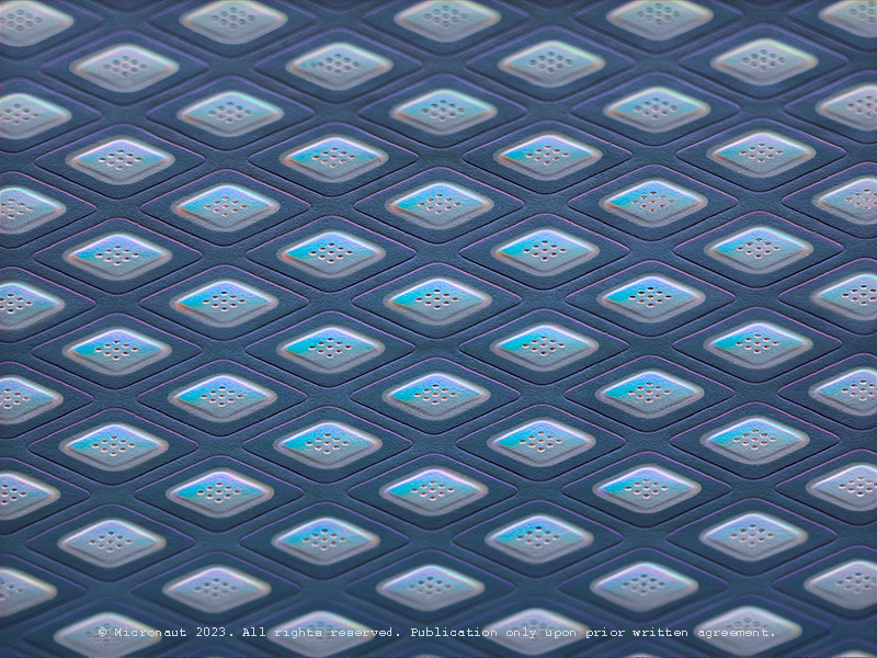
High-Density Microelectrode Array
This is a CMOS-based sensor-system designed to analyse neuronal signalling, drug discovery and cell assessment. It is equipped with 26,400 electrodes and developed to function within cell-culture incubators. Every human thought starts with a signal traveling from one neuron to another in the brain. Yet we know relatively little about how these connections form and grow into the highly ordered neuronal circuits, thereby connecting billions of cells in the brain of a human adult. Understanding how neural circuits are formed is one of the fundamental questions in neuroscience. To study the growth characteristics and signalling, scientists a semiconductor chip on which they can grow and analyze cells in realtime. This artwork was created by Martin Oeggerli (Micronaut). The picture is based on scanning-electron-microscopy technology. Colors are manually added by the artist in post-production to highlight the delicate interactions between brain cells.
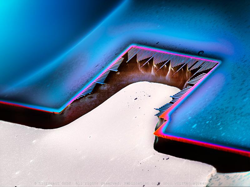
The Resonator
Hybrid spin-mechanical systems, formed by single spins coupled to mechanical resonators, have attracted ever-increasing attention over the past few years, triggered largely by the prospect of employing such devices as high-performance nanoscale sensors or transducers in quantum computing networks. In the Quantum Sensing Group of Patrick Maletinsky at the University of Basel, such hybrid systems comprising diamond mechanical cantilevers with embedded defect center spins are investigated as depicted in the image. Thereby crystal strain occurring upon cantilever displacement affords a natural and intrinsic mechanism to couple both systems. The research ultimately aims at investigating the potential of these systems for future fundamental physics studies and applications in sensing or information processing.
Porcine retina (Sus domesticus)
Porcine Retina (Sus domesticus) With far more than 100 million nerve cells, the retina is our first stage of the visual system and window to the outside world. The picture highlights the neuronal layers of a porcine retina. Seungkuk Ahn and his colleagues have electromagnetically guided viruses to specifically transduce each of the retina layers. The process inside the retina includes detection of light impulses (photons) by different light receptors, in general called rods (120 million cells) and cones (6 million cells), and the fast and continuous translation, filtration and post-procession into electrical signals (or nerve impulses). These signals are then passed through the optical nerve’s 1.5 million fibres to the visual centre of the brain and reinterpreted into a cohesive image.The flexibility and economy with which retinal cells work together remains beyond our powers of imagination: the eye reports numerous signals to the brain at once, including separate detection of light (light on) and dark (light off), general patterns and finest details, movements and different hues (including: red, green and blue). This literally makes the eye a camera with 10 to 15 different films and despite it appears so naturally to us, the perception of an image is the result of a most complex interaction process involving millions of cells and the electrical signals they produce and pass on to the brain.
Pheaodactylum tricornutum
Pheaodactylum tricornutum