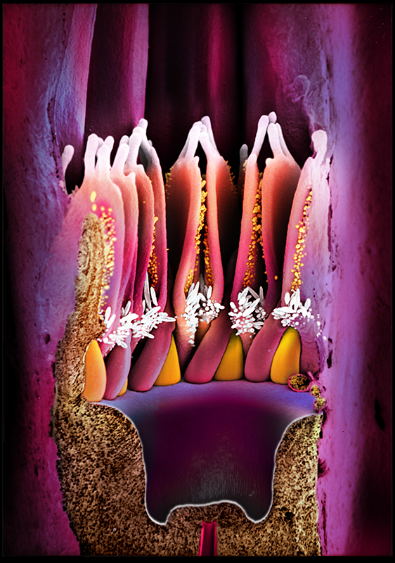
Microcosm II (plants & fungi)
This is the place of the earth’s smallest living organisms and their structures: concealed by sheer size, various forms of life spend their just below the radar of the human eye. Micronaut collects and explores invisibly small creatures and their structures from every corner of the earth. All images are based on Scanning-Electron-Microscopy (SEM). By utilizing electrons instead of photons, SEM technology is capable to produce magnifications up to 500.000-times or even more, thereby expanding the limits of traditional photography.
To reconstruct the colors -which cannot be captured through SEM technology- the pictures are manually colored (post-processing). The manual work combines Dr. Oeggerli’s scientific knowledge and his artistic eye, creating new insights into scientific phenomena and unknown territory. The following two projects contain selected images of plants/fungi.
«The images you have provided for the exhibition are looking excellent. Our whole design team has been blown away by them.»
Johanna Simkin
Senior Curator, Human Biology & Medicine, Museums Victoria
Micronaut images are rights-managed. If you want to get a quote, please contact us, providing the following information: (1) image name, (2) specific use, (3) industry, (4) distribution area, (5) format, (6) circulation or print run, and (7) duration.
Please note that we cannot answer incomplete requests. Thank you.
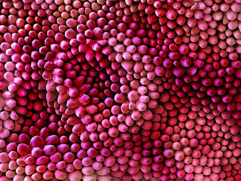
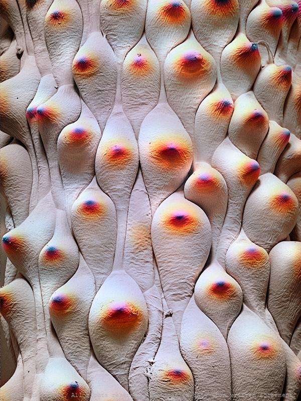
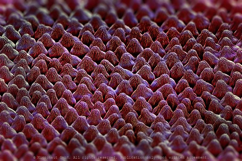
Velvet Underground (Rosa sp.)
Velvet UndergroundColored Scanning Electron Micrograph of the fine structures of a Red rose petal (Rosa sp.). Hairs are only present on the lower surface of the leaf. Thousands of finely corrugated warts cause the velvet shimmer on the upper surface of dark red rose petals.A superhydrophobic surface is a surface on which a drop of water forms an almost perfect sphere and even a very slight tilting is sufficient to cause the water drop to roll off. Biological tiny structures have been observed on many kinds of surfaces such as lotus leaves, rice leaves, butterfly wings, mosquito eyes, moth eyes, cicada wings, gecko feet, desert beetle, spider silks, fish scales, and red rose petals which exhibit excellent hydrophobicity and/or superhydrophobicity. Understanding the anti-wetting principles of surfaces is of special interest, because properties such as anti-sticking, anti-contamination, and self-cleaning are expected, and therefore surfaces with superhydrophobic properties are attractive for many industrial and biological applications, such as anti-biofouling paints for boats, anti-sticking and self-cleaning windshields and windows, microfluidics, stain resistant textiles, anti-soiling architectural coatings, or dust-free coatings on building glasses, and other biological and technical applications.
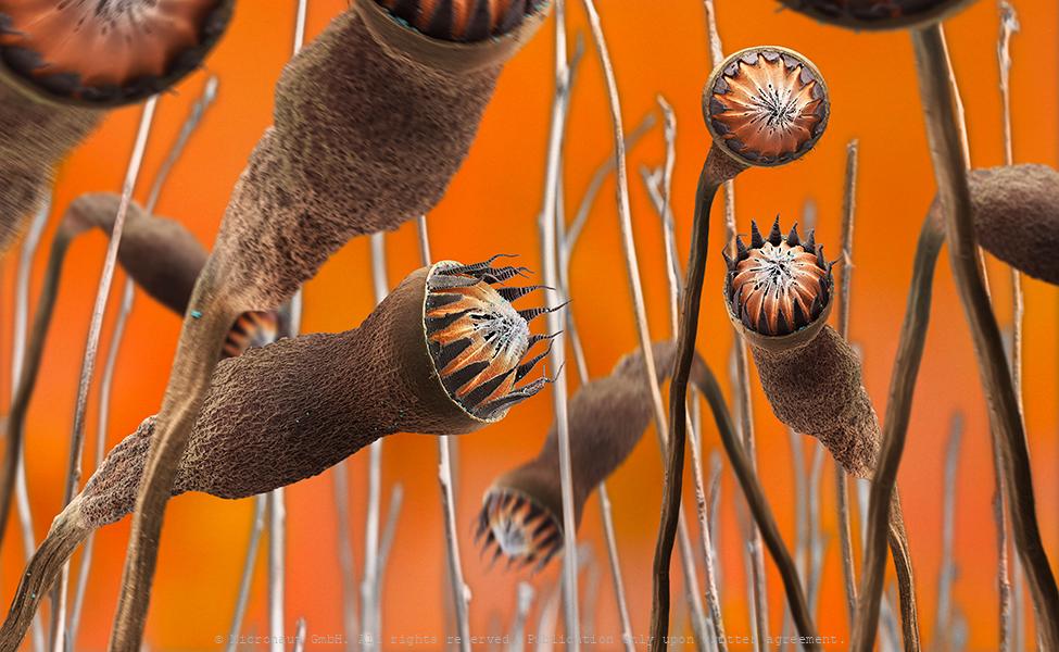
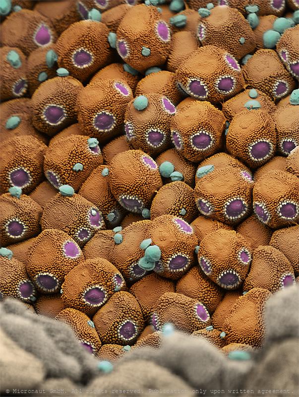
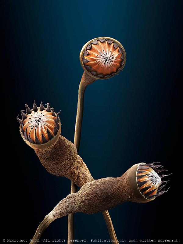
Dry moss capsules (Bryopsida sp)
Land plants have life cycles in which a haploid gemetophyte generation alternates with a diploid sporophyte. In mosses (Bryopsida sp.), the gametophyte is the dominant generation which is rarely the case in other species, and the sporophyte grows from the tip of a gametophyte. Sporocytes consist of a sporangium and a stalk. The Sporangium (or capsule) contains thousands of tiny spores which are released and distributed by the wind, after the lid has sprung open. This is the worlds largest color Scanning-Electron-Micrograph. The original print is measuring 250cm x 150cm (100 x 60in) and available in a limited edition of 3 pieces.
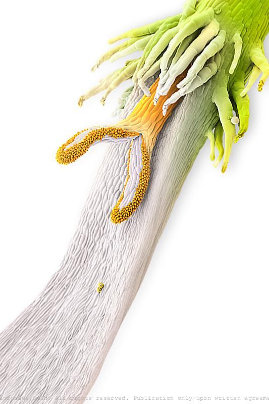
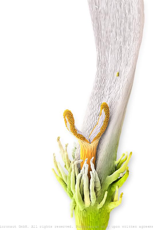
Floral Elements (Bruguiera gymnorhiza) - Nr.1
The species is Bruguiera gymnorhiza produces red pendulous flowers which attract birds (hence, bird pollination). This picture shows a longitudinal section through a young flower: at the very bottom you can see the "basal wing" of a young petal that guides a young stamen inside the petal. Next the curved stamen will elongate upwards. Reference page: http://iriomote.image.coocan.jp/research/mangrove/Bruguiera/BruguieraEnglish.html
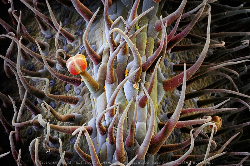
Pitcher plant (Nepenthes hemsleyana), Nr.1
Hand colored scanning-electron micrograph by Martin Oeggerli (Micronaut), showing the slippery surface inside a pitcher plant. Nepenthes hemsleyana is a tropical pitcher plant endemic to Borneo, where it grows in peat swamp forest and heath forest. It appears to rely on a prey trapping strategy unlike other pitcher plants: Interestingly, N. hemsleyana seems to have co-evolved with Hardwicke's woolly bats (Kerivoula hardwickii) which commonly roost in the upper pitchers. Studies shows that the carnivorous plant attracts bats by possessing modified pitfall taps that increase the reflectivity of echolocation calls - bats benefit by finding roosting sites, and the plants gain by receiving nitrogen from bat guano. Unlike in closely related pitcher plants, the upper pitchers of N. hemsleyana feature an expanded waxy zone (this structure is shown in the image) and a watery, less viscoelastic pitcher fluid. The pitchers also appear to lack UV patterns and produce less nectar and odour attractants, which is not required to host the bats.
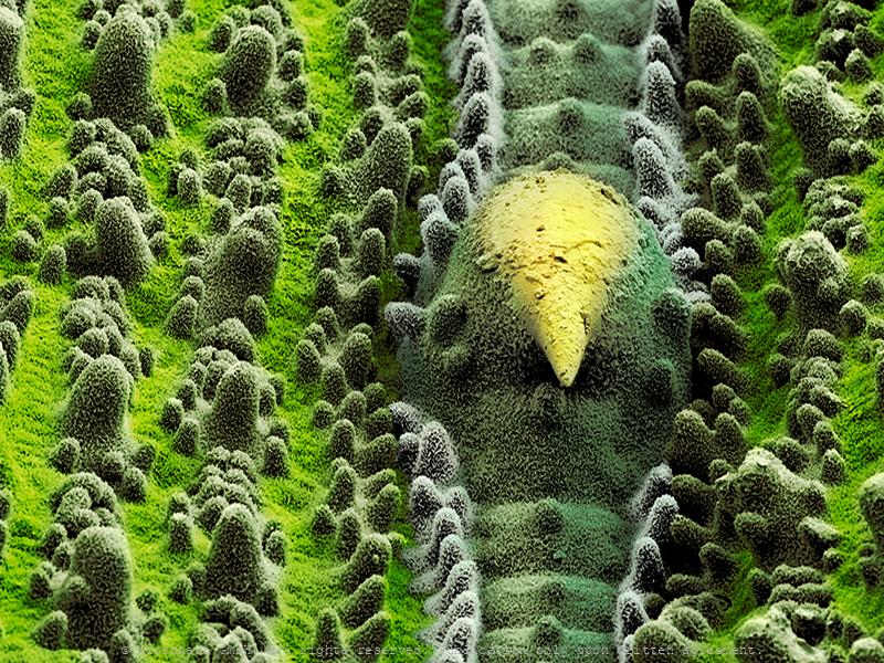
Martian Landscape (Loasa tricolor)
As protection for the ovaries, the gynoeceum wall of Loasa tricolor reveals similarities with a bouncy castle. Analysed under high magnification, many plant surfaces remind us on martian landscapes. Oeggerli slowly breathes life into his works by painstakingly selecting and masking different structures with color, layer upon layer utilizing his laptop touchpad. It is a slow manual process which allows him to set focus on mysterious microscopic structures which look almost extra-terrestrial.
Der Zauberpilz - The Psycodelic Mushroom (Claviceps purpurea)
Lysergic acid diethylamide is a synthetic lysergamide that can be obtained as a derivative of naturally occurring ergot alkaloids. LSD is one of the most powerful hallucinogens known and belongs to a subgroup of psychedelics that affect the body's serotonin system. In mid-April 1943, the Swiss chemist Albert Hofmann accidentally discovered the psychoactive substance LSD. The drug has since been banned, but in the right dosage and with therapeutic support, LSD can also be used as a helpful medication against depression. Thinking, feeling and perception are massively influenced by the psychoactive substance, the experience of space and time changes, sensory illusions, colors, image distortions or delusions may occur. -- Lysergsäurediethylamid ist ein synthetisches Lysergamid, das als Derivat natürlich vorkommender Mutterkornalkaloiden erhalten werden kann. LSD ist eines der stärksten bekannten Halluzinogene und gehört zu deren Teilgruppe der Psychedelika, welche auf das Serotonin-System des Körpers wirken. Mitte April 1943 entdeckte der Schweizer Chemiker Albert Hofmann per Zufall die psychoaktive Substanz LSD. Zwischenzeitlich wurde die Droge verboten, doch in richtiger Dosierung und in therapeutischer Begleitung kann LSD auch als hilfreiches Medikament gegen Depressionen eingesetzt werden. Denken, Fühlen und die Wahrnehmung werden durch die psychoaktive Substanz massiv beeinflusst, das Erleben von Raum und Zeit verändert sich, es kann zu Sinnestäuschungen, bunten Farben, Bildverzerrungen und Wahnvorstellungen kommen.
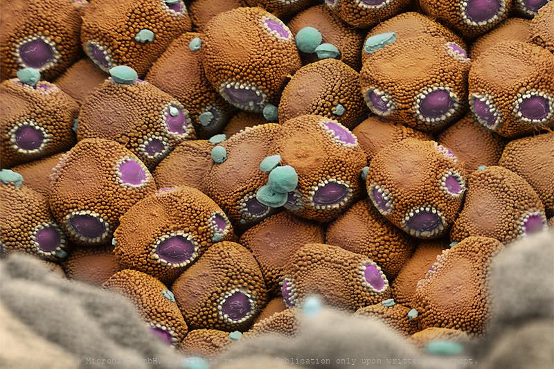
Rust fungus spores
hese fungal spores inside the fungal fruitbody of a Rust fungus (Urediniomycetes sp.) wait to be dispersed by the wind. Image was awarded 'Best Scientific Cover Image 2008'.
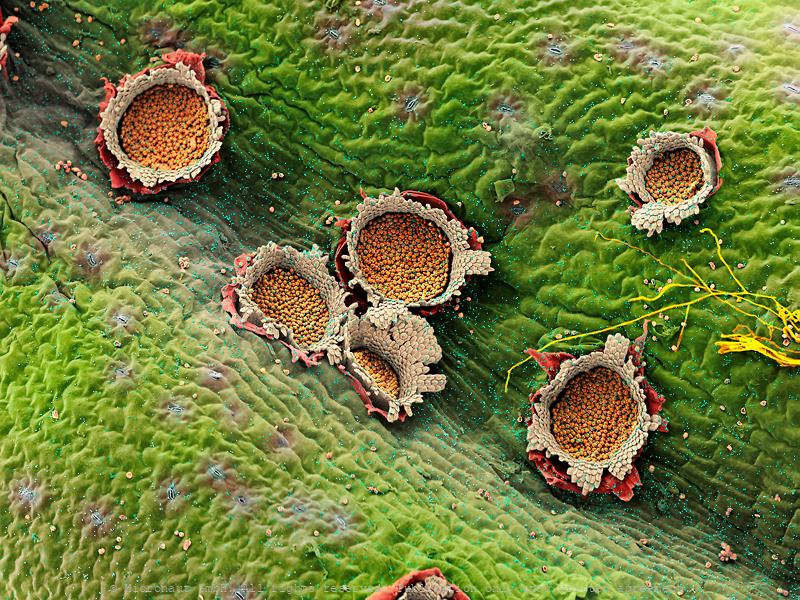
Rust fungus fruitbodies
The image shows a plant which has been infected by a Rust fungus. After reproduction of the fungus, it produces fruitbodies which are perforating through the epidermal layer of the plant leaf to release the tiny spores. Rust fungus are a dangerous threat for monocultures, e.g. soy and wheat.
Rust fungus spores
hese fungal spores inside the fungal fruitbody of a Rust fungus (Urediniomycetes sp.) wait to be dispersed by the wind. Image was awarded 'Best Scientific Cover Image 2008'.
Germinating Fungus Spore (Original of Evil)
Artwork created by Martin Oeggerli (Micronaut). The picture is based on scanning-electron-microscopy technology, and all colors are manually added by the artist in post-production. Urera baccifera is a species of flowering plant in the nettle family known by many common names, including scratchbush, ortiga brava, pringamoza, mala mujer, chichicaste, nigua, guaritoto, ishanga, manman guêpes, and urtiga bronca. The tree can reach five meters in height. The thin, toothed leaves are up to 25 centimeters long and are covered by stinging hairs. On this leaf, a fungi spore rests under a bulbous gland. The parasite has already started to grow hyphae, eventually trying to invade the leaf.
Fungal attack
Fungi spore sitting and waiting in front of a plant pore (Wheat)
A Coral in the Forest (Nectria cinnabarina)
Nectria cinnabarina, also known as coral spot, is a plant pathogen that causes cankers on broadleaf trees. This disease is polycyclic and infects trees in the cool temperate regions of the Northern Hemisphere. N. cinnabarina is typically saprophytic, but will act as a weak parasite if presented with an opportunity via wounds in the tree or other stressors that weaken the tree’s defense to the disease. Drought, other fugi and physical damage can make a tree susceptible to this pathogen which allows infection and leads to pink fungal blobs (indicative of its sexual stage) on the outside of dead wood. The pathogen thrives in dead wood and airborne spores infect living trees and shrubs through wounds. In summer and autumn, orange-red fruiting structures are produced; eventually these structures mature to dark red and can survive through the winter. This asexual stage is characterized by spongy conidia which can be distinguished by the hard, dark red blobs on the bark. Both of these structures release spores that can be dispersed by water and invade susceptible tissue. Nectria cinnabarina was first described in 1791 belongs to the same family as far better known Fusarium oxysporum f.sp. cubense, which represents a big threat for banana plants worldwide, causing the Panama disease.
Ticket to Heaven - Pollen on a Flower Petal (Barringtonia sp.)
Their size is measured in millionths of a meter, but the romantic journeys of pollen are epic: a spherical shaped medium sized pollen grain takes a rest before continuing the voyage and eventually become the first - and only - that achieves fertilization. Coloured scanning electron micrograph (SEM) of a Putat tree (Barringtonia asiatica) pollen grain on a flower petal. Artwork by Martin Oeggerli (Micronaut), 2021.
Spotlessly clean - Self cleaning properties of plants
Biological surfaces are usually not flat. Instead, most plants reveal complex three dimensional structures down to the nanometer range and often look highly ordered. It's an amazingly beautiful microscopic world. But what are the reasons behind? Plants are unable to move. To remain clean, they have to rely on self cleaning properties. Over millions of years, plants have developed sophisticated repellent structures, which also make sure leaf- and flower surfaces do not stick together, not even under wet circumstaces. In addition, flowers start as small buds and often unfurl immensly large petals within a short period of time. This extreme volume changes require special tissues that are both flexible and tear-resistant - similar to a parachute. In fact there are so many funtional reasons for the fascinating diversity of microscopic plant surfaces that it is impossible to mention all of them. Beyond that, nature often finds more than one solution to solve a problem. It only adds to the complexity for those trying to explore and understand the vast diversity of plant micro-structures.
The self-cleaning flower petal (Agrostemma githago)
Surfaces define boundaries and interactions between different surfaces, including an individual and its outside world. Surfaces also play crucial roles in entire ecosystems and are of particular importance for plants which are sessile organisms with large functional surfaces: e.g. due to large grasslands, it can be estimated that 250 million km2 of superhydrophobic leaf surfaces exist on eartch, which equals about 50% of the earth’s total surface. Over several hundred million years, plants have developped a variety of different functional surfaces, including velvety leaves which have self-cleaning properties. This image shows the petal of a common corn-cockle (Agrostemma githago) under high magnification. The flower is pink-to-white and the epidermis contains hundreds of convex microstructures.
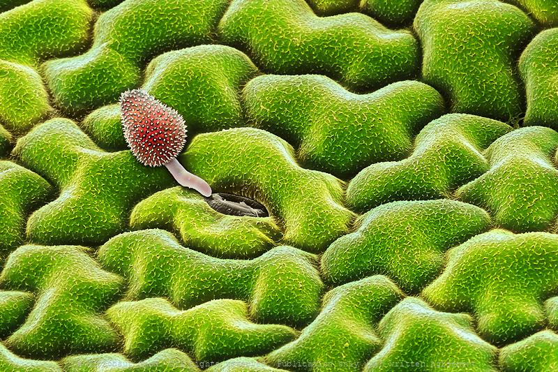
Soja rust spore
Coloured scanning electron micrograph (SEM) of a Rust fungus spore on a freash Soja leaf. The germinating fungi spore grows towards a plant pore (stomata), to infect its host plant. Rusts are parasitic fungi that cause some of the most destructive plant diseases. The disease appears as numerous rusty, orange spots that rupture the epidermal layers of the leaves. The fungus rarely causes the plant to die but greatly reduces the ability to photosynthesize (produce food from sunlight), often leading to severely reduced yields.
Nightshade stars (Solanum pyrocanthos)
Nightshade stars - Star-shaped prickles, developing from furry trichomes, densely coat the leaves and stems of the Porcupine tomato. The Porcupine Tomato, Solanum pyrocanthos, belongs to a diverse and cosmopolitan family of plants (Solanaceae) with over 1,500 members including the tomato, potato and nightshades. The plant contains various toxic tropane alkaloids in its leaves, stem and fruit and therefore should be considered dangerous to humans. Strong and straight fluorescent orange thorns occupy the stems and leaves of the plant (not shown on image), giving it a foreboding appearance. The prickles are consistent throughout the plant, developing clearly from furry trichomes which densely coat the plant's leaves (on both sides) and stems.
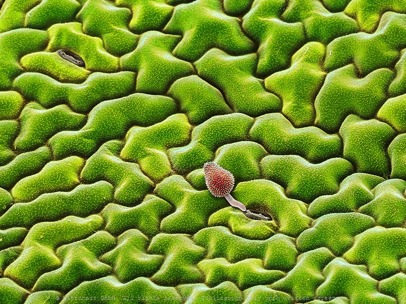
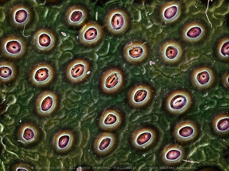
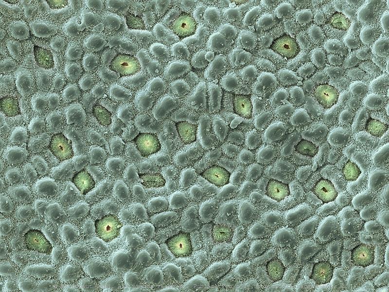
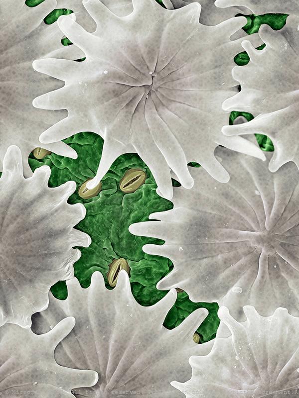
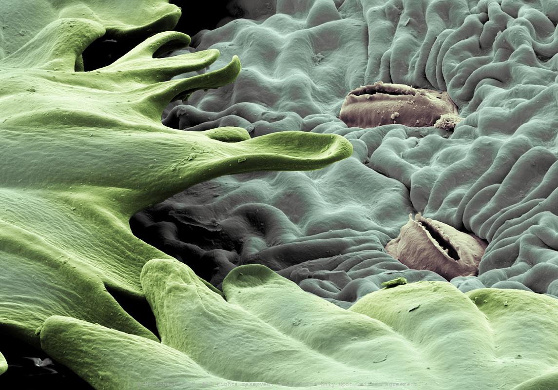
Green Earth - Leaf of a Fig Tree (Ficus benjamini)
Plants "breath" through pores, or stoma, on the lower surface of their leafs. Each stoma can be opened or closed through regulations of the turgor pressure of the surrounding guard cells. Thereby, plants can take in carbon dioxide and release oxygen. Stomata are tiny, microscopic and needed for photosynthesis. Thousands of them dot on the surface of the plants. Each pore resembles a doughnut and consists of two cells - each known as a guard cell. They can swell or shrink to open or close the pore which allows the plant to regulate gas exchange for photosynthesis and control moisture levels in tissues depending on inner and outer stimuli. This picture shows the aggromerated stomata on the lower surface of Ficus benjamini. The picture is based on a scanning electron micrograph and was hand-colored by science artist Martin Oeggerli.
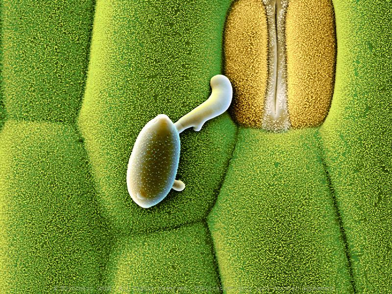
Keep breathing - Plant epidermis with open pore (called stoma)
Plants "breath" through stoma on the lower surface of their leafs. Each stoma can be opened or closed through regulations of the turgor pressure.
Ilex leaf (Ilex sp.), Nr.2
Plants "breath" through stoma on the lower surface of their leafs. Each stoma can be opened or closed through regulations of the turgor pressure.
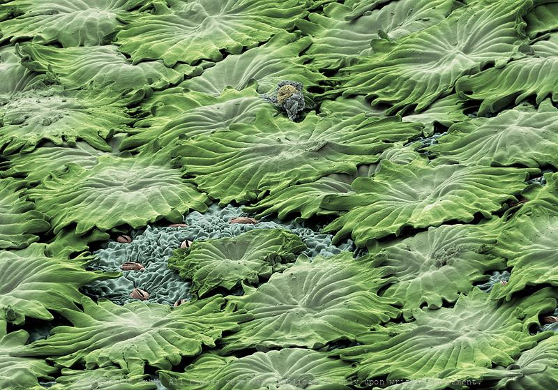
Grass surface with pores, Nr. 1
Grass surface with pores, Nr. 1 (green)
Grass surface with pores, Nr. 2
Grass surface with pores, Nr. 2 (closed)
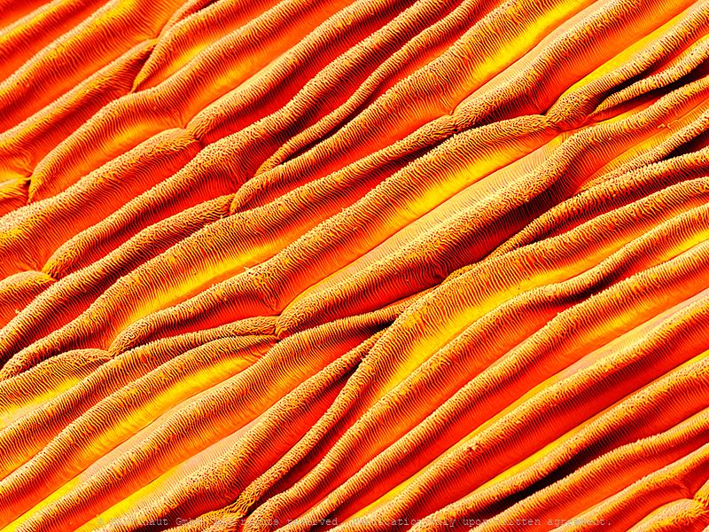
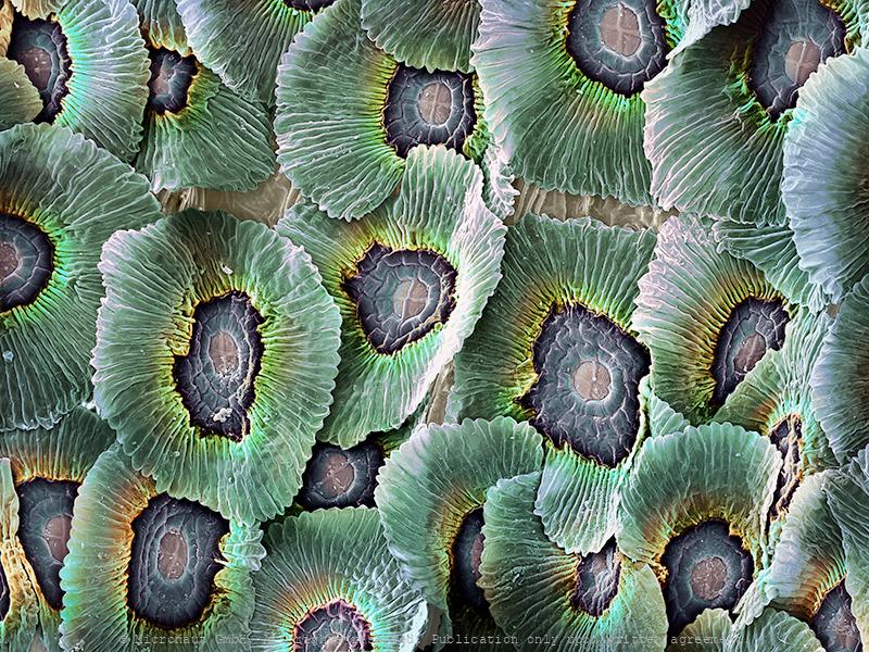
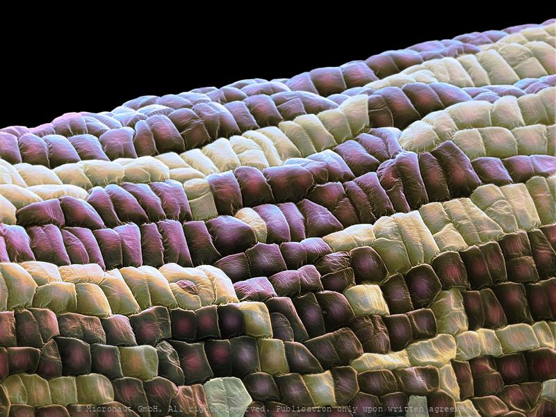
Rose Petal Surface, Nr.1
Lower surface of a Rose petal
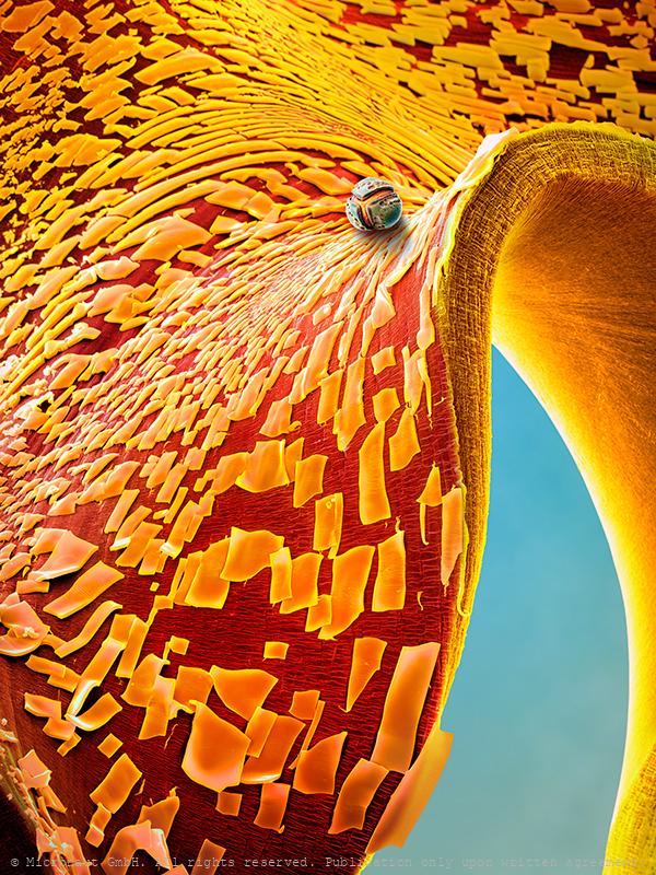
Velvet Underground - Rose petal Nr.2
Velvet UndergroundColored Scanning Electron Micrograph of the fine structures of a Red rose petal (Rosa sp.). Hairs are only present on the lower surface of the leaf. Thousands of finely corrugated warts cause the velvet shimmer on the upper surface of dark red rose petals.A superhydrophobic surface is a surface on which a drop of water forms an almost perfect sphere and even a very slight tilting is sufficient to cause the water drop to roll off. Biological tiny structures have been observed on many kinds of surfaces such as lotus leaves, rice leaves, butterfly wings, mosquito eyes, moth eyes, cicada wings, gecko feet, desert beetle, spider silks, fish scales, and red rose petals which exhibit excellent hydrophobicity and/or superhydrophobicity. Understanding the anti-wetting principles of surfaces is of special interest, because properties such as anti-sticking, anti-contamination, and self-cleaning are expected, and therefore surfaces with superhydrophobic properties are attractive for many industrial and biological applications, such as anti-biofouling paints for boats, anti-sticking and self-cleaning windshields and windows, microfluidics, stain resistant textiles, anti-soiling architectural coatings, or dust-free coatings on building glasses, and other biological and technical applications.
Eucalyptus leaf area
Eucalyptus leaf area
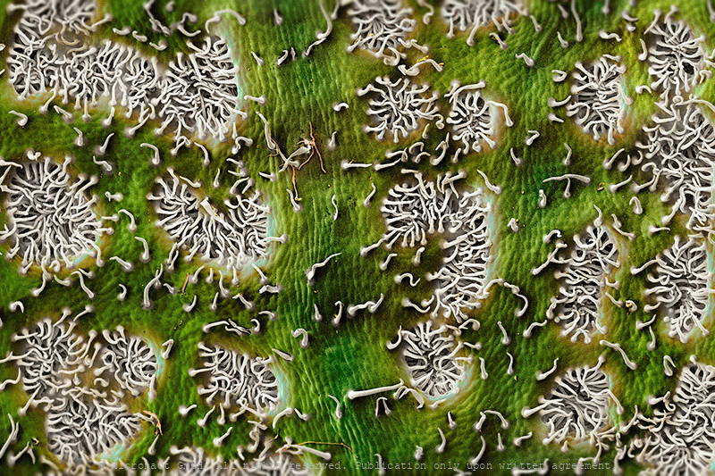
Peaceful Wilderness - The Oleander Leaf (Nerium oleander)
Under the powerful magnification of the scanning-electron-microscope (SEM), familiar objects turn into alien landscapes: this picture shows an ordinary oleander (Nerium oleander) leaf which is an every-day plant that can be found across the mediterranean and also throughout northern Europe, due to an increasing popularity among gardeners. While contemporary natural landscapes have been colonized and domesticized on a global scale over the past decades, this peaceful place on an Oleander leaf appears to have completely escaped contamination by human intervention - despite actually being part of it. The plant grows in a small bead & breakfast lodge close to the city of Basel, Switzerland.
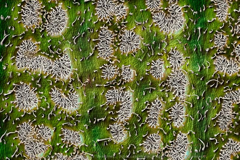
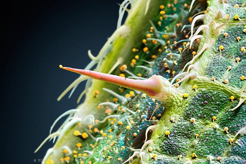
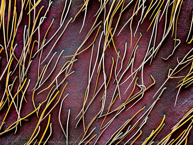
Stinging Nettle hair (Urtica dioica)
The Stinging Nettle (Urtica dioica) is a plant with soft green leafs that are equipped with glands as well as short non-stinging and special elongated stinging hairs (center), from which the species derives its common names: stinging nettle, burn nettle, burn weed, or burn hazel. Stinging hairs (trichomes) produce a painful stinging sensation by injecting a chemical mixture when touched by humans or other animals. They act like hypodermic needles: after the tip breaks off, a chemical mixture composed of histamine, acetylcholine, 5-HT (serotonin), moroidin, leukotrienes and formic acid is injected and causes pain or paresthesia. Furthermore, the plant has a long history of use in medicine, as food source (tea) and as source of fibre.
Leaf surface - Magnification makes you a stranger
Surface of a leaf with hairs
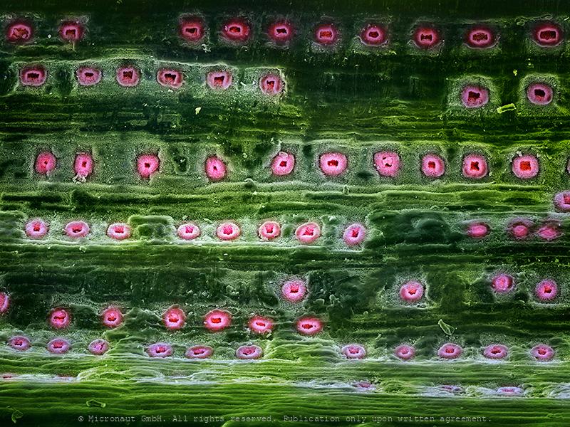
Conifer needle epidermis
Conifers have needles instead of leafs, which are also green and photosynthetically active. Depending on the species they persist 1.5-40 years and must be much more stably built than leafs of deciduous trees.
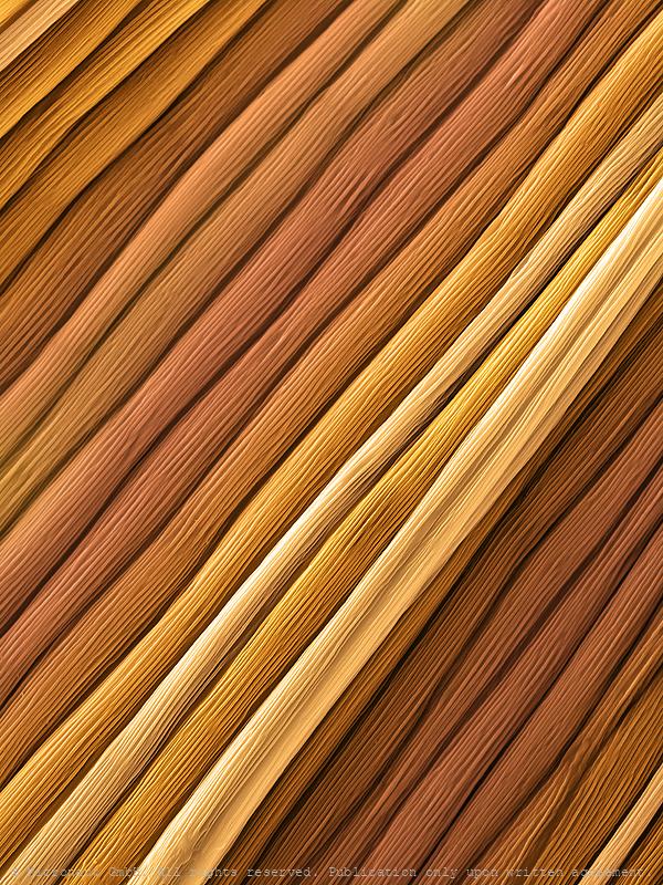
Walnut stem (Juglans niger), Nr.1
Drüsenhaare auf Walnuss Epidermis, trichomes on walnut epidermis
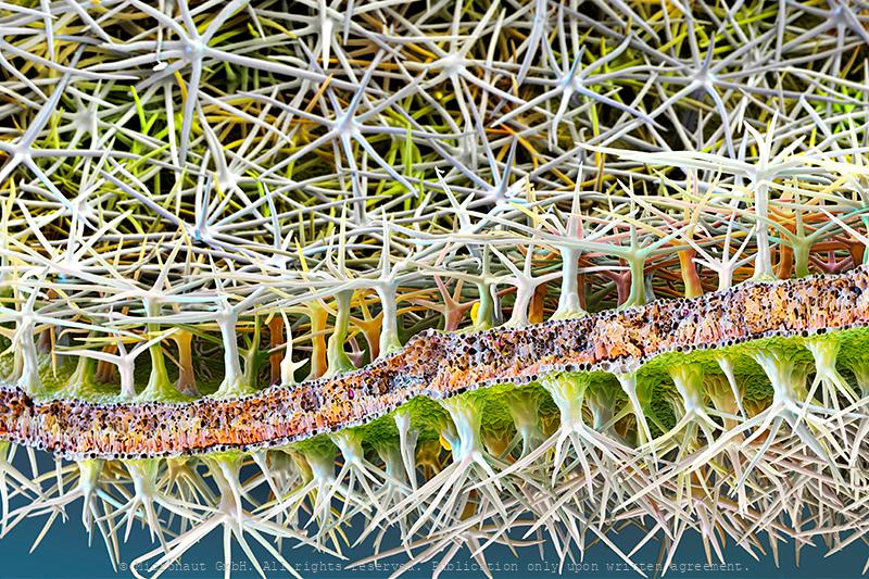
The epidermis of a tillandsia is covered with sucking hairs. The plant has reduced roots and is specialised on using fog as the primary source of water.
As water becomes a more and more scarce, it's interesting to look around and learn how nature is dealing with the exact same problem, and it reveals interesting techniques and structures. While some plant species such as e.g. Eucalyptus are covering the entire leaf area with a thick layer of wax, the Olive tree has developed flattened hairs on the leaf surface which resemble umbrellas to provide some extra shade for the leaf surface below. Yet the Tillandsia went one step further: the small epiphytic plant which has no roots and virtually no access to groundwater resources, uses special trichomes (which are called sucking hairs) to absorb water from moist air. The picture shows how trichomes cover the entire leaf area. It needs a unique solution to survive in an acrobatic environment.
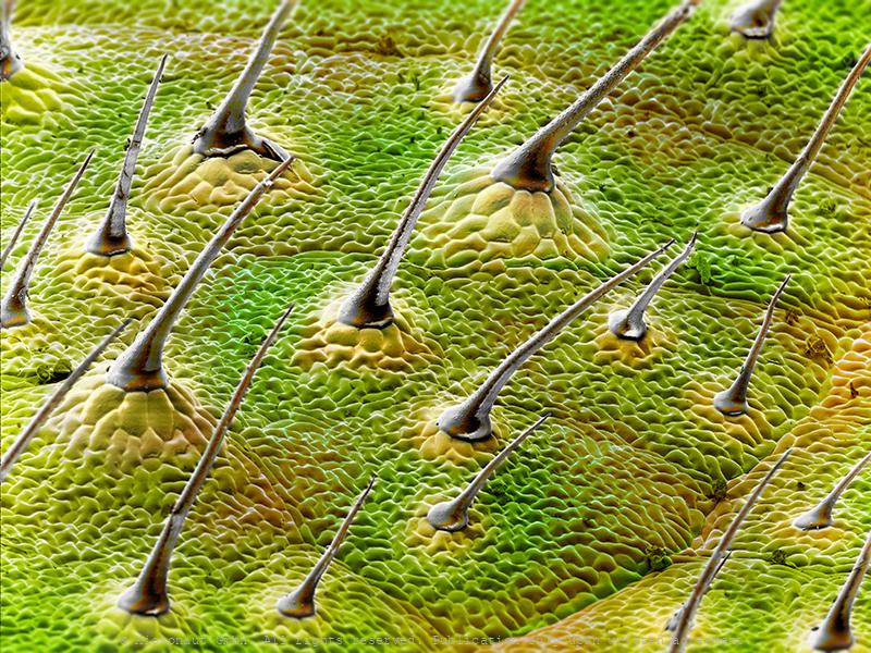
Olive leaf (Olea europaea), Nr.3
The surface of the olive tree leaf is covered by trichomes (hairs that resemble umbrellas). The function of the trichomes is to provide shade and safe water. They can also recycle water, which is lost during transpiration through the stoma.
Olive leaf (Olea europaea), Nr.1
Olive leaf (Olea europaea), Nr.2

Germinating Fungus Spore (Original of Evil)
Artwork created by Martin Oeggerli (Micronaut). The picture is based on scanning-electron-microscopy technology, and all colors are manually added by the artist in post-production. Urera baccifera is a species of flowering plant in the nettle family known by many common names, including scratchbush, ortiga brava, pringamoza, mala mujer, chichicaste, nigua, guaritoto, ishanga, manman guêpes, and urtiga bronca. The tree can reach five meters in height. The thin, toothed leaves are up to 25 centimeters long and are covered by stinging hairs. On this leaf, a fungi spore rests under a bulbous gland. The parasite has already started to grow hyphae, eventually trying to invade the leaf.
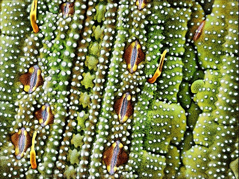
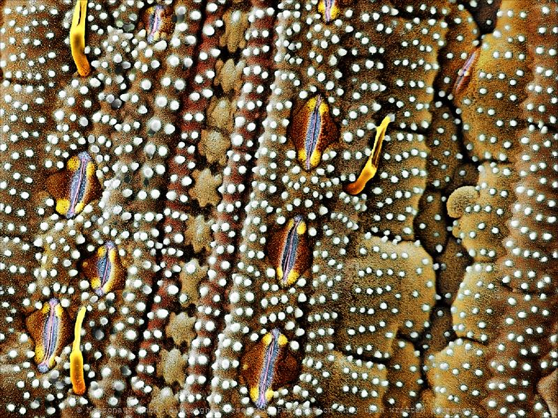
Tomato leaf (Lycopersicon esculentum)
Tomato leaf surface. Hand-colored scanning electron micrograph (SEM) of the surface of a tomato leaf (Lycopersicon esculentum) created by Martin Oeggerli. The picture is showing the complexity of a plant leaf area at invisibly small dimension. Scientists believe that the hairs and glands protect the plant against predators and reduce water loss through evaporation. Also visible are discretely grey-blue colored pores (stomata). They are responsible for the gaseous exchange: CO2 enters the leaf, while O2 and water are evaporated, thereby maintaining a constant flow of water from the roots, through the stem and branches, to the leafs. Glandular trichomes are a special type of 'hairs' and contain crystals and oils in the bulbous section of the structures. Two of these structures can be seen in the picture. It’s believed that the crystals and oils are part of the plant’s defense mechanisms. The essential oils are responsible for giving the tomato plant its characteristic smell. The volatile compounds of the oils that contribute that contribute to the typical scent of tomato leafs are (Z)-3-hexenal, limonene, hexanal, (E)-2-hexenal, eugenol, 1,8-cineole, caryophyllene, beta-phellandrene, humulene, and linalool. It’s a little ironic that what might be considered intoxicating to us can be so “unpleasant” to pests! Collectively, these compounds are exclusive to tomato trichomes.
Rice leaf (Poaceae sp.)
superhydrophobic leaf surface of a rice leaf with spike
Waterrepellent micro-surface (Salvinia molesta)
Aquatic organisms possess structured surfaces which enable them to effectively retain air films while submerged in water. The transfer of these optimised hydrophobic structures on technological surfaces would open up a multitude of applications.
©-Micronaut-Gynoecium-Surface-PTU0031_mini_wet
Flower landscape (Erica sp.)
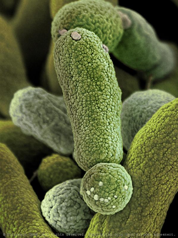
Aster pollen grains
Petal surface (Salix caprea)
Daisy leaf (Bellis perennis)
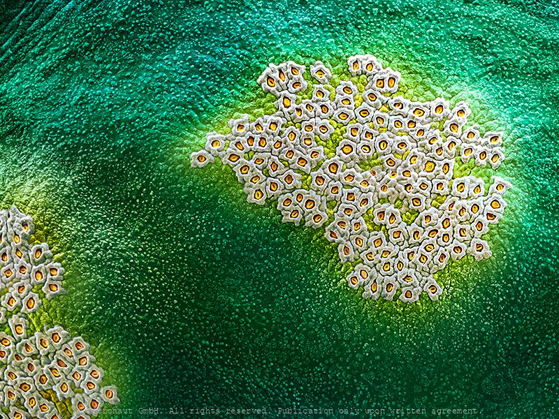
Biological surfaces are usually not smooth. They mostly possess complex three dimensional structures, down to the nanometer range. Flowering plants have evolved structured surfaces which are optimised to facilitate the attachment of insects.
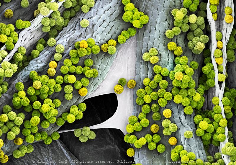
Ragweed pollen (Ambrosia artemisiifolia)
Ragweed, a rather unremarkable plant holds a stupefying record: it produces 1.6 million highly allergic pollen per hour, thereby knocking-out hayfever sufferers all around the world...

Fern leaf (Sori & Spores)
Fern Leaf with Sori and Spores (and plant pores; stomata). About 12,000 species of plants belong to the ferns, an ancient botanical group of plants known as Pteridophyta. Unlike mosses, they have xylem and phloem, thus they are vascular plants. They have stems, leaves, and roots like other vascular plants. Ferns reproduce via spores and have neither seeds nor flowers. The life cycle of ferns is different from flowering plants and characterized by alternating diploid sporophytic and haploid gametophytic phases. The gametophyte of ferns is a free-living organism, whereas the gametophyte of the gymnosperms and angiosperms is dependent on the sporophyte.
Rhizobium sp. Nr.1
Rhizobium sp. highly enlarged. This bacteria species is used for the biosynthesis fo L-carnitine
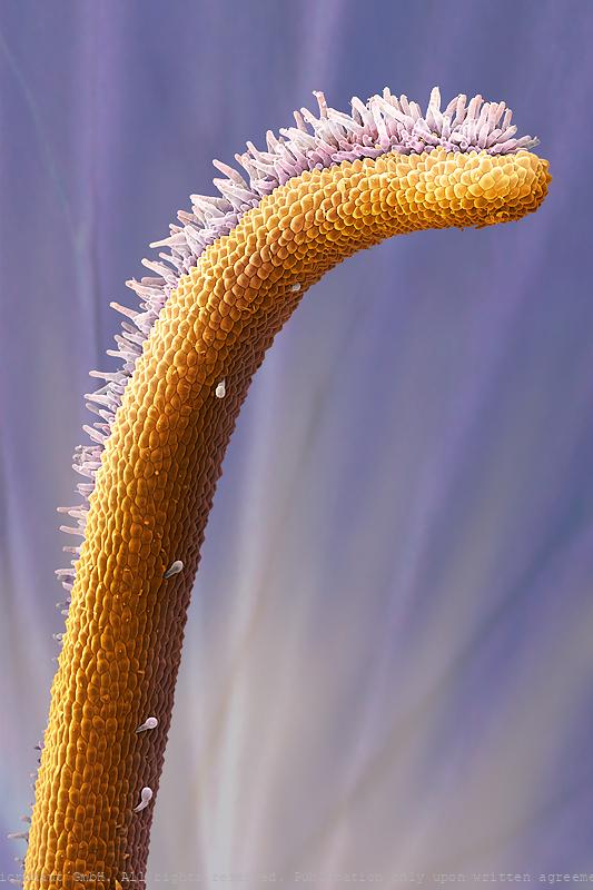
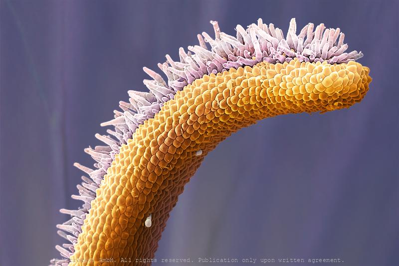
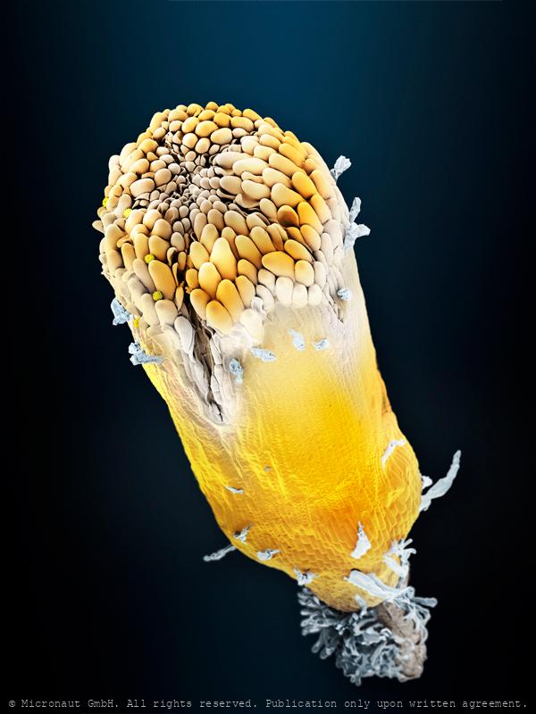
Pistil, Nr.3
A pistil is formed from the stigma (tip), style (elongated part) and the ovary (bottom part; not visible). Small pollen (male reproductive cells) reache the female ovary by attaching on the stigma, growing inside the style and entering the ovule.
Pistil, Nr.3
A pistil is formed from the stigma (tip), style (elongated part) and the ovary (bottom part; not visible). Small pollen (male reproductive cells) reache the female ovary by attaching on the stigma, growing inside the style and entering the ovule.
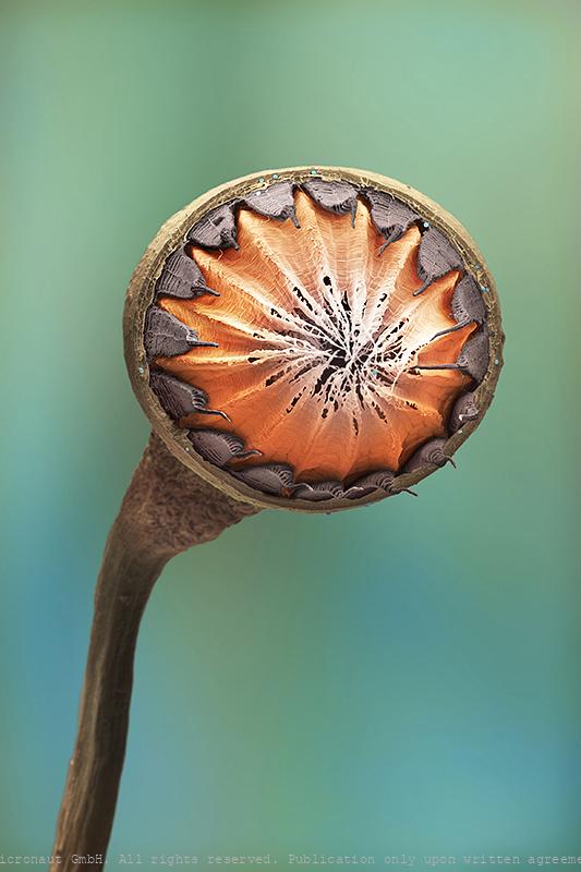
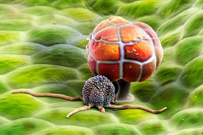
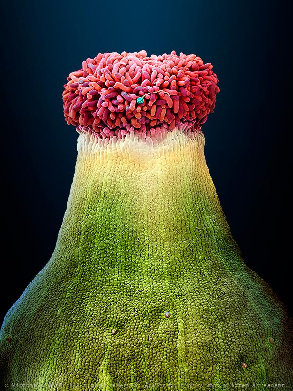
Dry moss capsule (Bryopsida sp)
dry moss capsule from the front view. after extensive drought, the capsule explodes and sets free the tiny spores (thousands of them). the elongated "stick", terminating into the capsule, will make sure all spores are distributed easily by the wind.
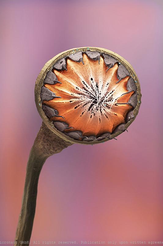
Dry moss capsule (Bryopsida sp)
dry moss capsule from the front view. after extensive drought, the capsule explodes and sets free the tiny spores (thousands of them). the elongated "stick", terminating into the capsule, will make sure all spores are distributed easily by the wind.
Dry moss capsules (Natura obscura)
dry moss capsule from the front view. after extensive drought, the capsule explodes and sets free the tiny spores (thousands of them). the elongated "stick", terminating into the capsule, will make sure all spores are distributed easily by the wind.
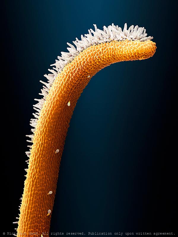
Saffron pistil (Crocus sativus)
Saffron (Crocus sativus) pistil; manually colored 3D realistic scanning electron micrograph by Martin Oeggerli / Micronaut, 2018. Saffron is a spice derived from the flower of Crocus sativus, commonly known as the “saffron crocus”. The vivid crimson stigmas and styles, called threads, are collected and dried to be used mainly as a seasoning and colouring agent in food. Saffron, long among the world’s most costly spices by weight, was probably first cultivated in or near Greece. Its recorded history is attested in a 7th-century BC and it has been traded and used for over four millennia. Iran now accounts for approximately 90% of the world production of saffron.Saffron’s taste and iodoform or hay-like fragrance result from the chemicals picrocrocin and safranal. It also contains a carotenoidpigment, crocin, which imparts a rich golden-yellow hue to dishes and textiles. Saffron contains more than 150 volatile and aroma-yielding compounds. It also has many nonvolatile active components, many of which are carotenoids, including zeaxanthin, lycopene, and various α- and β-carotenes. However, saffron’s golden yellow-orange colour is primarily the result of α-crocin.
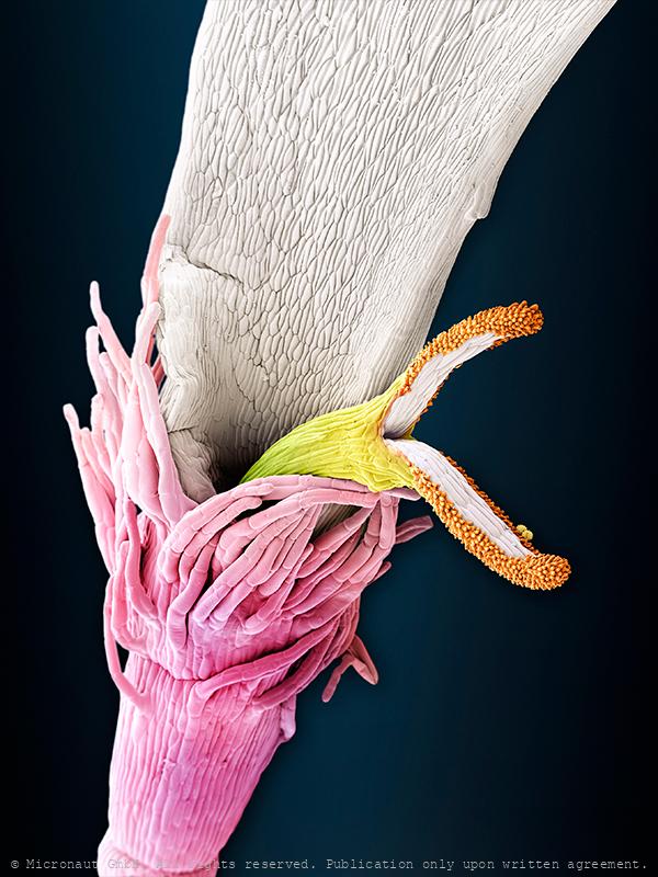
Erotic Adventures of the little Chardonnay pollen (Vitis sp.)
Pistil of a Chardonnay flower, including a pollen grain -placed in the central part of the stigma- that is growing a pollen tube. Chardonnay is a green-skinned grape variety used in the production of white wine. The variety originated in the Burgundy wine region of eastern France, but is now growing wherever wine is produced, from England to New Zealand. The Chardonnay grape itself is very neutral, with many of the flavors commonly associated with the grape being derived from such influences as terroir and oak. Chardonnay is also an important component of many sparkling wines, including Champagne. Modern DNA fingerprinting research suggests that Chardonnay is the result of a cross between the Pinot noir and Gouais blanc. The Romans are thought to have brought Gouais blanc from Croatia, and it was widely cultivated by peasants in eastern France. The Pinot of the French aristocracy grew in close proximity to the Gouais blanc, giving both grapes ample opportunity to interbreed.
Pistil, Nr.3
A pistil is formed from the stigma (tip), style (elongated part) and the ovary (bottom part; not visible). Small pollen (male reproductive cells) reache the female ovary by attaching on the stigma, growing inside the style and entering the ovule.
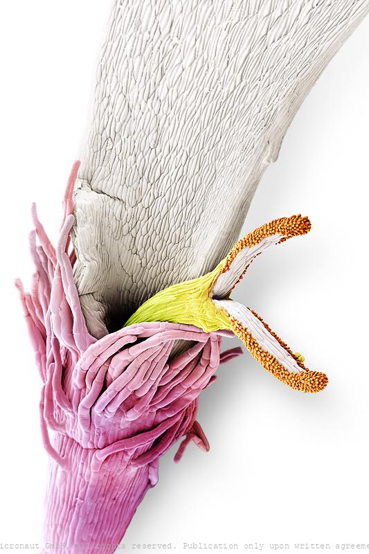
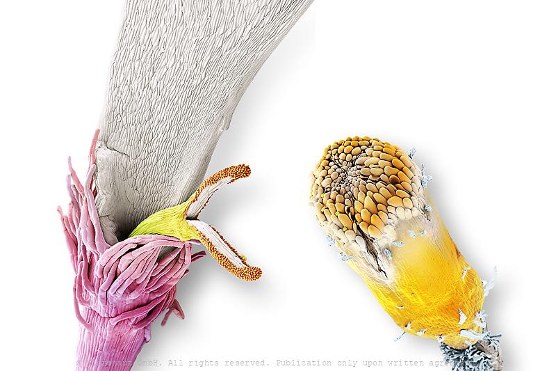
Peripheral floret from a Composite Flower (B)
Single floret from a compound flower (The Common Daisy; Bellis perennis) with two small pollen sticking to the tip of the pistil. Florets from the border of the influorescence have developped a special morphology with five long clean and seamlessly adnate white sepals, in order to attract pollinators.
Daisy floret (Bellis perennis), Nr.2
Single floret from a compound flower (The Common Daisy; Bellis perennis). Florets from the center of the influorescence have developped a special morphology with completely reduced sepals.
Peripheral floret from a Composite Flower (A)
Single floret from a compound flower (The Common Daisy; Bellis perennis) with two pollen sticking to the white petal. Florets from the border of the influorescence have developped a special morphology with five long clean and seamlessly adnate white sepals, in order to attract pollinators.
Peripheral floret from a Composite Flower (B)
Single floret from a compound flower (The Common Daisy; Bellis perennis) with two small pollen sticking to the tip of the pistil. Florets from the border of the influorescence have developped a special morphology with five long clean and seamlessly adnate white sepals, in order to attract pollinators.
Peripheral floret from a Composite Flower (A)
Single floret from a compound flower (The Common Daisy; Bellis perennis) with two pollen sticking to the white petal. Florets from the border of the influorescence have developped a special morphology with five long clean and seamlessly adnate white sepals, in order to attract pollinators.
Comparison of two flower morphologies present in Asteraceae
Comparison of two florets from a compound flower (The Common Daisy; Bellis perennis). Florets from the border of the influorescence (left) have developped a special morphology with five long clean and seamlessly adnate white sepals, in order to attract pollinators, whereas florets from the center have totally reduced sepals. Central florets are densely packed and mainly required for increased pollen- and seed production.
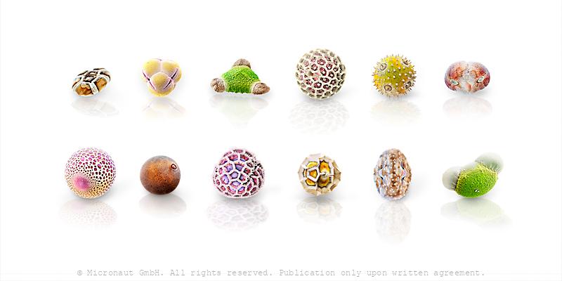
The universe of pollen
Pollen grains are remarkably diverse and vary in size from 10 to 250 µm. First illustrations of pollen grains start in the 17th century, shortly after the development of the first light microscope. At the end of the 19th century most characteristics of the capsule structure have already been established. One and a half centuries later, invention and commercialization of scanning-electron-microscopy (SEM) allow fascinating new insights into the microcosmos and re-initiate pollen research. The hand-colored SEM images of pollen that have been assembled on this picture illustrate the fascinating diversity of these small carriers of the male plant genome.