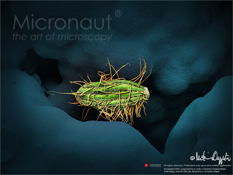
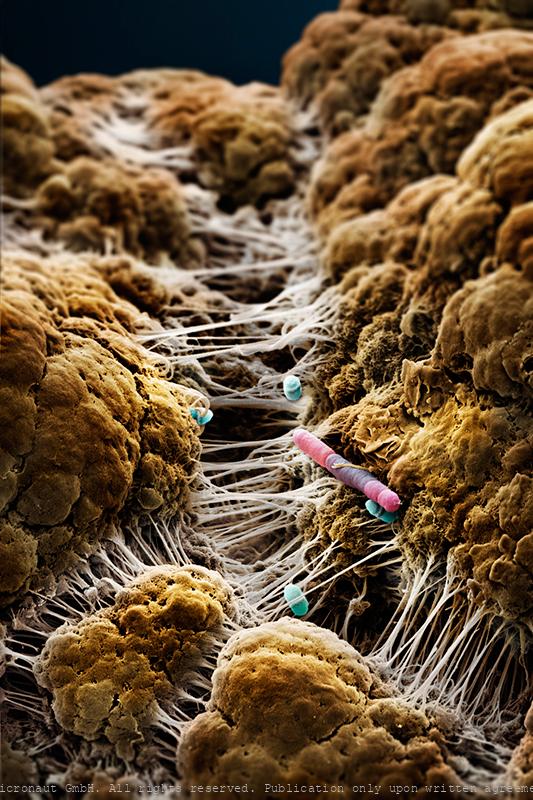
CHASING MICROBES
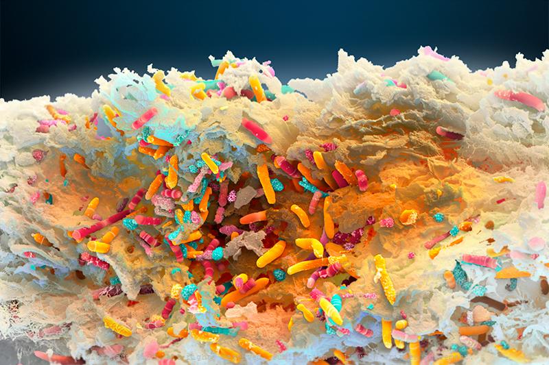
The human gut microbiome flaunts its diversity in this sample of human feces, which includes an enormous bacterium that’s about 50 times longer than E. coli. Everyone’s mix of microbes is unique. Microbiome research is relatively young, and scientists are still learning the many ways these microbes affect our health, weight,
mood, and even personalities. In the not-too-distant future it might be routine to deliver a dose of healthy microbes in the form of prebiotics, probiotics, or fecal transplants—helping us realize the promise of operating at top form, from the inside out.
«The images are utterly sensational! The clarity and colors are gorgeous… truly one of a kind images.»
Shannon Hibberd
Senior Photo Editor, National Geographic
Micronaut images are rights-managed. If you want to get a quote, please contact us, providing the following information: (1) image name, (2) specific use, (3) industry, (4) distribution area, (5) format, (6) circulation or print run, and (7) duration.
Please note that we cannot answer incomplete requests. Thank you.
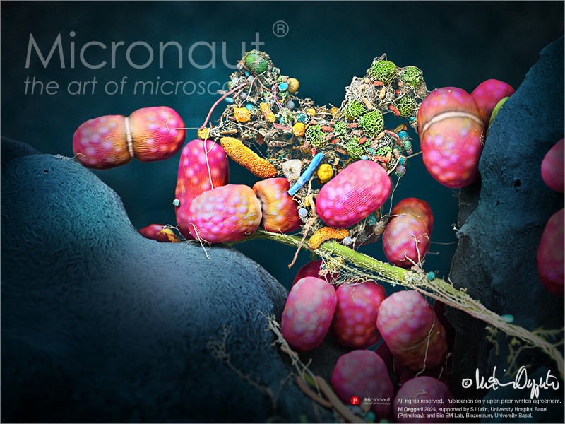
human Microbiome of a KISS
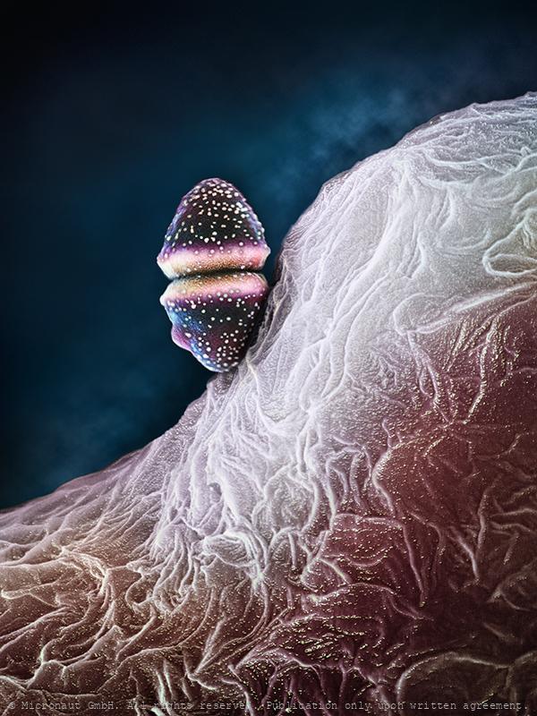
The Human Microbiome of a Kiss. Artwork created by Martin Oeggerli (Micronaut) and dedicated to his 8-years old son Nelson. This image was originally produced for the National Geographic feature article 'How trillions of microbes affect every stage of our life-from birth to old age'. Soon after its public release, our daily life became heavily overshadowed by the world's coronavirus (COVID-19) pandemic - thereby instantly confirming the articles visionary title. Meanwhile (at end of April 2020), the crisis has not only severely limited the artists ability to work, but also prohibits him from officially visiting his son who is living in Switzerland, just a stone's throw away from the rest of the father's German-based patch-work family. This picture is based on scanning-electron-microscopy technology, and all colors are manually added by the artist in post-production, to visualize the immense diversity of the microbial community transmitted through a kiss. Whenever we kiss someone on the lips or cheeks invisibly small microbes are exchanged from one person to the other one! The variety of microbes that are part of our microbiome is mind-blowing, and the composition of the invisibly small communities can be the cause of many factors, including the human genetic makeup, diet, age, surroundings, and sexual behavior. Among humans, approximately 90% of cultures have some type of kissing. Usually it is platonic, such as a parent kissing a child. However, in 77 of 168 (46%) of all cultures, it can go as far as intimate (French-) kissing. Recently, a scientific study has revealed that on average 80 million bacteria are transferred to the partner during a kiss of 10 s. Most partners share a more similar oral microbiome compared to unrelated individuals and/or e.g. their kids. But some of the collective bacteria among partners are only transiently present, while others have found a true niche and survive permanently, allowing long-term colonization.With regard to kissing, th
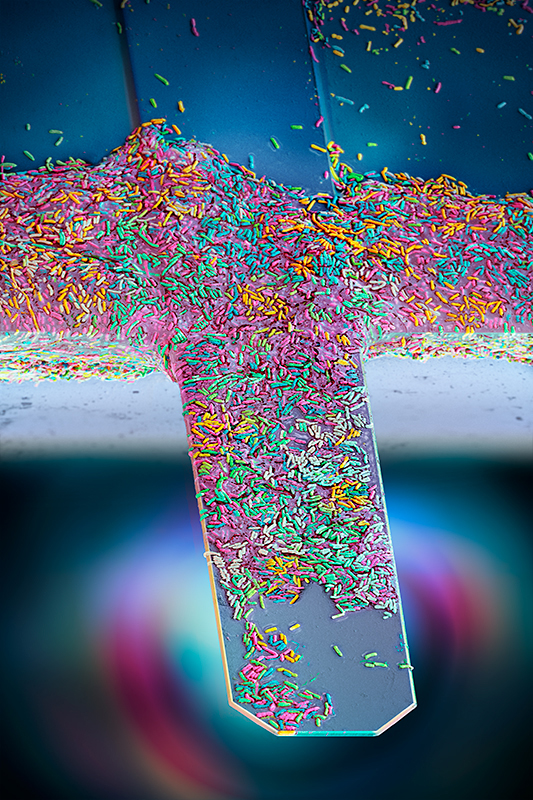
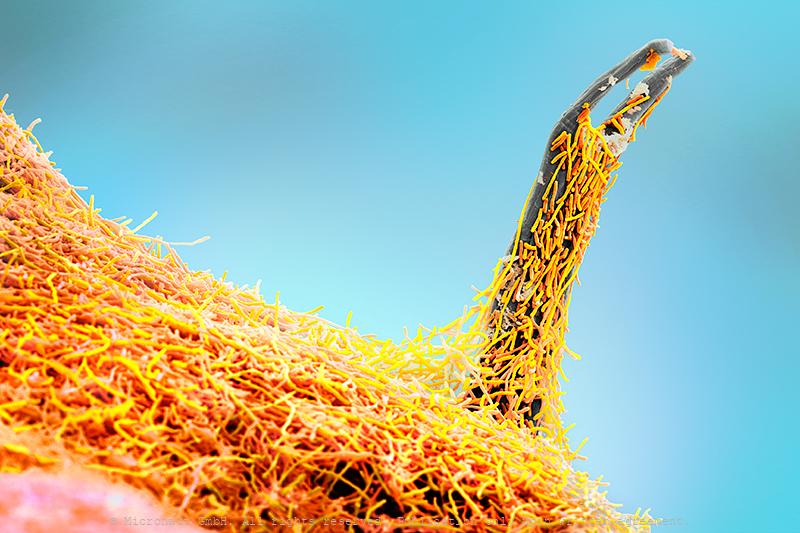
Nanomotion sensor - detecting the vibration of living bacteria i
Antibiotic Resistance Chip Loaded with Bacteria. Hand-colored SEM scan by Martin Oeggerli / Micronaut, 2021. Antibiotic resistance is currently one of biggest threats to global health. By offering a diagnostic device, Resistell proposes an alternative to culture based antibiogram, the current gold standard in antibiotic susceptibility testing. The rapid AST method is based on the detection of movement caused by living bacterial cells. Because the test is growth independent rapid AST, we reduce the time taken to get a result from days to minutes. Resistell provides information on which antibiotic should be used to treat the patient, and the concentration at which it should be administered. This rapid AST designed and developed by Resistell saves lives by finding the right antibiotic on time and reduces spreading of antibiotic resistance!
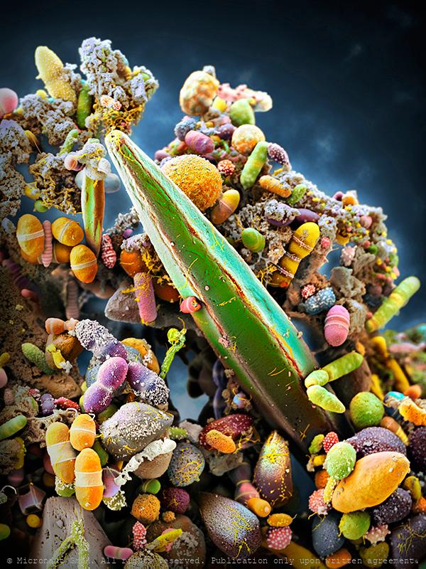
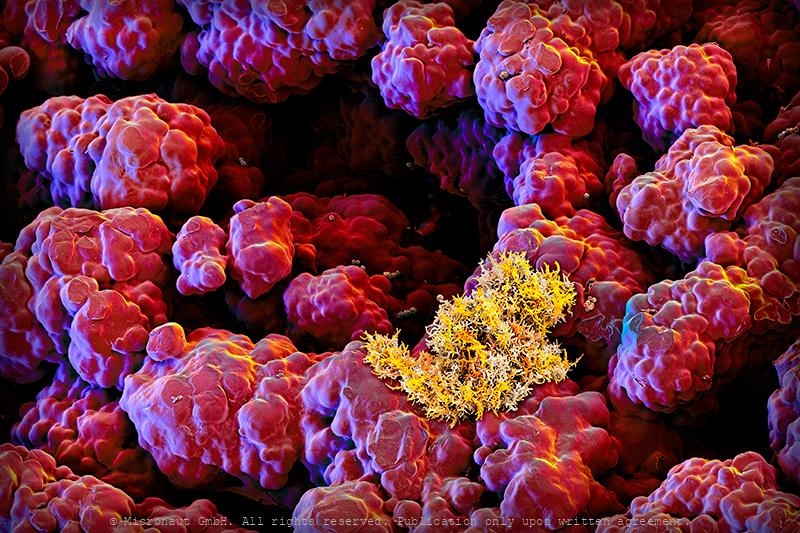
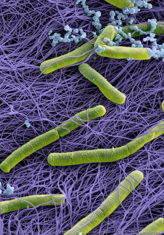
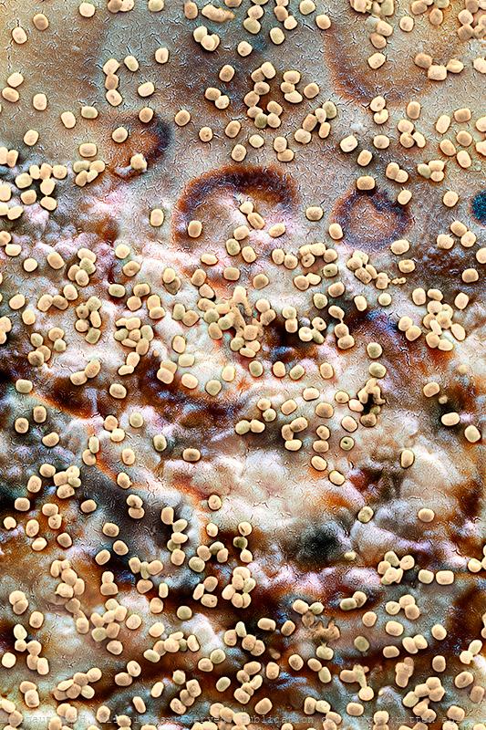
The Human Microbiome, Nr.1
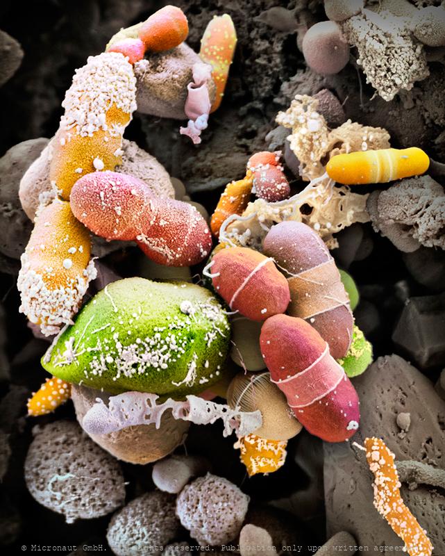
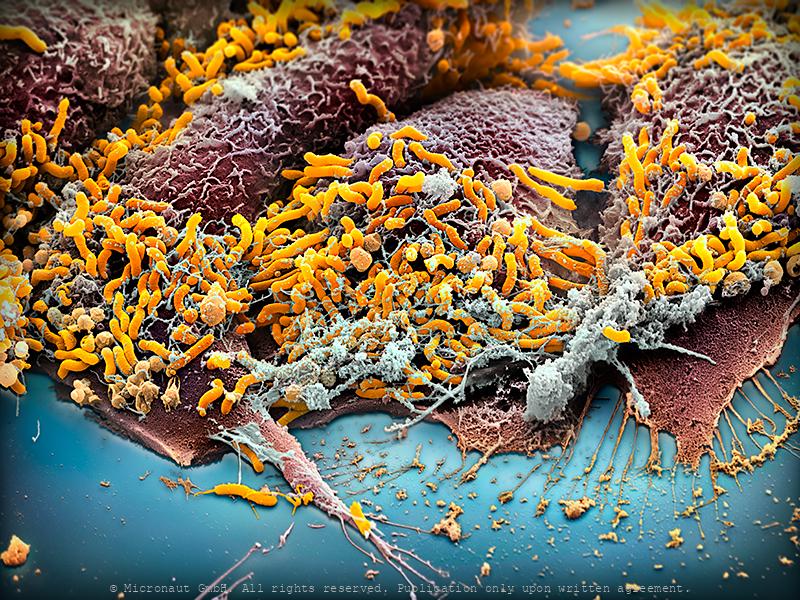
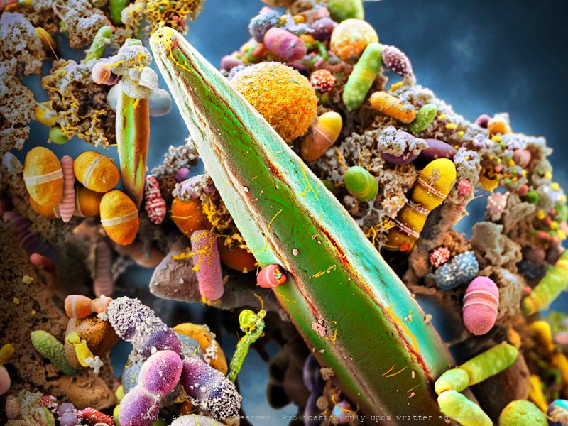
Artistic coloration of human excrements, that contain billions of intestinal bacteria, including Epulopiscium sp. - the world’s largest bacteria species! Artistic coloration of the microbiome present in a healthy human gut, containing billions of intestinal bacteria, including Epulopiscium sp. - the world’s largest bacteria species! The human gut teems with bacteria: streptococci, staphylococci, enterococci, enterobacteria, mycobacteria, spirochetes, mycoplasma, corynebacteria, clostridia, and lactobacilli. Many of their species are still unknown. They help us digest food and absorb nutrients, or play a role in protection of intestinal walls. Many gut bacteria further regulate weight and ward off autoimmune diseases. In adult humans, the intestinal flora is responsible for up to five percent of the body weight, forming a fragile ecosystem (the human microbiome) which is sensitive to: stress, unhealthy food, alcohol or illness. Under unfavorable circumstances our health is directly affected by bacteria that live inside of us, and as a result we may suffer from: overweight, mood changes or become prone to various diseases. In the central part of the image you can see the world’s largest bacteria - Epulopiscium sp. which spans between 200 – 700 µm, i.e. 350-times bigger than E. coli, or B. subtilis. Epulopiscium was first discovered in 1985 in the intestines of a surgeonfish, where it is only active during the day and believed to control the pH within the gut of its hosts. Because of its huge size, the giant exhibits many unusual characteristics, including a very special ‘rhomboid’ morphology. This image has been produced using scanning electron microscopy (SEM) in combination with selectively assigning different colors to different bacteria species and structures. By creating a 3D-like photorealistic appearence, the artist aims to expose the diversity of bacteria that can be found within human feces.
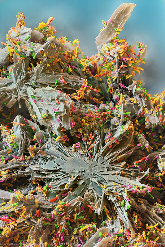
The Gut Microbiota Miracle - Microbiome of a Newborn (H. sapiens
The microbiome of a baby, 5-6 month after birth. Hand colored scanning electron micrograph by Martin Oeggerli (Micronaut). The Gut Microbiota Miracle: this picture shows the microbiome of a newborn one month after birth. The importance of our intestinal bacteria is already well known – but much less is known about its development and diversity from a newborn to the adult. It has long been assumed that human breast milk is sterile. But scientists have discovered complex and dynamic bacterial ecosystems in human breast milk which are distinct from other microbioal communities and may also colonize and widen the diversity of the infant gut probiotic bacterial community. According to the study, the cocktail of beneficial bacteria passed from mother to infant through breast milk changes significantly over time and could act like a daily booster for infant immunity and metabolism, with important implications for infant development and health. Bacteria found in breast milk include Actinobacteria, Bacillariaphyta, Bacteroidetes, Cyanobacteria, Deinococcus-thermus, Firmicutes, Fusobacteria, Patescibacteria, Plantae, and Proteobacteria.
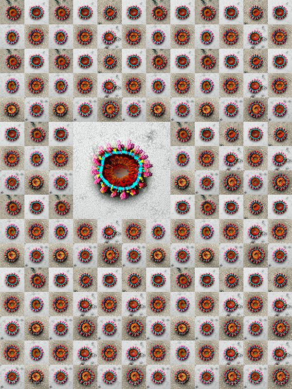
Ciliata
Ciliate from Cadagno Lake that has dips into the oxygen-free chemocline zone (where oxygen ends and a layer of anaerobic green and pink bacteria forms) to feed on anoxygenic microorganisms. The ciliates are a group of protists characterized by the presence of hair-like organelles called cilia (identical in structure to eukaryotic flagella, but shorter and present in much larger numbers). Cilia are used for swimming, crawling, attachment, feeding, and sensation. Ciliates reproduce asexually, by various kinds of fission and exchange DNA through conjugation. The fossil record goes back to 580 million years (Triassic period). Ciliates are commen almost anywhere in lakes, ponds, oceans, rivers, and soils, including anoxic and oxygen-depleted habitats. About 4,500 unique free-living species have been described - including many ectosymbiontic and endosymbiontic species - but the potential number of existant species is estimated at 27,000–40,000. Most ciliates are heterotrophs and feed on smaller organisms, such as bacteria and algae, and detritus swept into the oral groove (mouth).
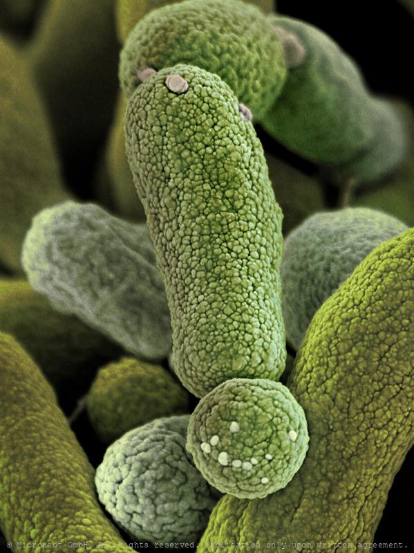
The Light Harvesting Bacterium (Chromatium okenii)
The Light Harvesting Bacterium (Chromatium okenii), Ehrenberg 1838, Perty 1852 Lake Cadagno is a very special aquatic system, showing a permanent stratification of the water throughout the year, with the upper oxygen-containing water layer above an anoxic layer rich in dissolved salts. Red sulphur anaerobic bacteria (RSA) of the species Chromatium okenii thrive at the interface of the two layers, where they are able to mix considerable volumes of water, not by stirring it directly with their flagella, but by clustering together in search of light in a restricted area near the oxygen diffusion front. In doing so, the volume density increases and the water begins to fall, taking the micro-organisms with it in a process known as bioconvection. Chromatium okenii are light sensitive anaerobic bacteria found in water. The cells are slightly curved or straight rods, measuring about 12-15 µm which is quite large for bacteria. Red sulfur bacteria contain chlorophyll a and carotenoids, both of which are located on the inner membrane of the sac type. The bacteria are capable of anoxygenic photosynthesis with oxidation of hydrogen sulfide to sulfur or sulfate (and so do not produce oxygen). In doing so, they accumulate sulfur in special compartments or granules (called 'sulfur balls') within the cell. The presence of photosynthetic pigments leads to various shades of orange to brown and pink to violet and purple to red. Strong growing populations of Red sulfur bacteria sometimes lead to red water layers and even entirely red lakes. In the case of this probe, the bacteria are also responsible for the detoxification of the aquatic system. This allows many more other organisms, including fish, to prosper in the relatively small alpine lake. Most of the organisms live in the layer near the surface (above the red water, inhabited by Chr. okenii). Below the red bacteria layer, a death zone extends towards the bottom of the lake. It is assumed that the anaerobic phototrophic sul
The Gut Microbiota Miracle - Microbiome of a Newborn (H. sapiens
The microbiome of a baby, 1 month after birth. Hand colored scanning electron micrograph by Martin Oeggerli (Micronaut). The Gut Microbiota Miracle: this picture shows the microbiome of a newborn one month after birth. The importance of our intestinal bacteria is already well known – but much less is known about its development and diversity from a newborn to the adult. It has long been assumed that human breast milk is sterile. But scientists have discovered complex and dynamic bacterial ecosystems in human breast milk which are distinct from other microbioal communities and may also colonize and widen the diversity of the infant gut probiotic bacterial community. According to the study, the cocktail of beneficial bacteria passed from mother to infant through breast milk changes significantly over time and could act like a daily booster for infant immunity and metabolism, with important implications for infant development and health. Bacteria found in breast milk include Actinobacteria, Bacillariaphyta, Bacteroidetes, Cyanobacteria, Deinococcus-thermus, Firmicutes, Fusobacteria, Patescibacteria, Plantae, and Proteobacteria.
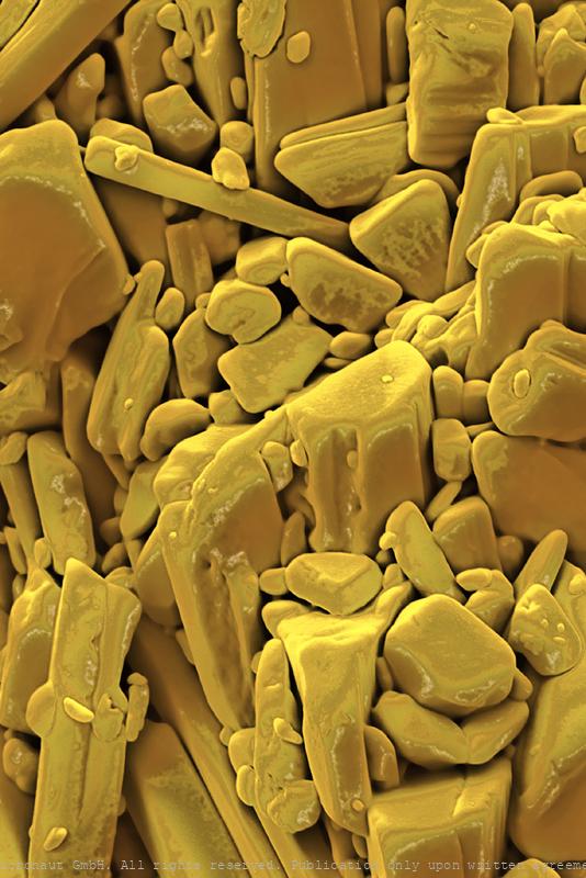
A Fuzzy Jacket for Cheese!
Artwork created by Martin Oeggerli (Micronaut). The picture is based on scanning-electron-microscopy technology, and all colors are manually added by the artist in post-production, to visualize characteristic microstructures of the microbiotic fungus Geotrichum candidum. The fungus G. candidum is a member of the human microbiome, notably associated with skin, sputum and feces where it occurs in 25–30% of specimens. It is also common in soil and has been isolated from soil probes collected across all continents. Furthermore, this yeast is the causative agent of geotrichosis. Pulmonary involvement is the most frequently reported form of the disease, but bronchial, oral, vaginal, cutaneous and alimentary infections have also been reported. G. candidum is also widely used in the production of certain dairy products including rind cheeses such as Camembert, Saint-Nectaire, Reblochon and other wrinkly cheeses that you may find at your local cheese shop. In a Nordic yogurt-like product known as viili, the yeast can be found as well, being responsible for the product's velvety texture. Furthermore, G. candidum has been reported as an organism that is able to break down plastic. Using in-depth genomic sequencing, French scientists recently unlocked the evolutionary history of this important cheese microbe and revealed a fungus with an identity crisis: all true yeasts are descendants of molds. They evolved a single-celled lifestyle from their moldy multi-cellular ancestors. Throughout this evolutionary process, they let go of habits such as growth using the filaments that create the fuzzy appearance of most molds. For a long time, Geotrichum candidum (hereafter G. candidum) has been a mycological mystery to both cheese makers and scientists. It has a fuzzy appearance when it grows on the surface of cheeses or on Petri dishes in the lab, but when you look under the microscope, it also has the appearance of tiny single cells that look like a yeast. And when scientists st
The Human Microbiome, Nr.2
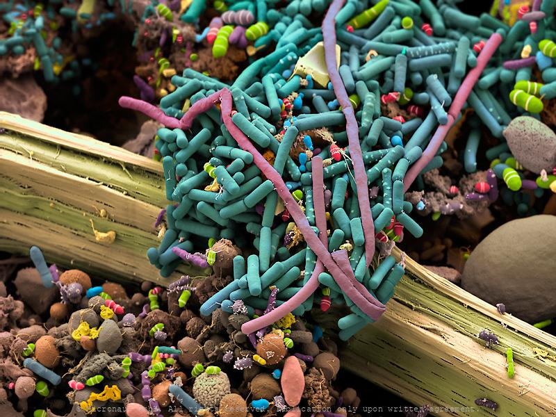
Excrements revealing the bacterial flore of the human intestinal tract. If it comes to digestion, we are not alone! Among many more bacterial forms streptococci, staphylococci, enterococci, enterobacteria, mycobacteria, spirochetes, mycoplasma, corynebacteria, clostridia, or lactobacilli are living in our gastro-intestinal tract and usually help us to digest food, aid nutrient absorption, reduce dehydration or assist in the production of key vitamins. In adults, the bacterial flore makes up about five percent of the total body weight (similarlly to the brain). Altogether, more than 30'000 different species of bacteria have been classified in our intestinal tract, so far! However, it’s a fragile ecosystem. The bacterial flore is sensitive to stress, unhealthy food, alcohol or illness. Under unfavorable circumstances it directly affects our health and e.g. causes overweight, changes our mood or makes us prone to various diseases. The small intestine normally contains relatively low numbers of bacteria compared to the large intestine. If abnormally large numbers of bacteria grow in the small intestine, they may use many of the nutrients that a person would normally absorb for their growth. Too much growth and breakdown of nutrients can damage the cells of the small intestinal wall, leading to Crohn’s disease, diabetes, scleroderma and severely influence the progression of AIDS or immunoglobulin deficiency. This hand-colored scanning electron micrograph reveals the large number and variability of bacteria that are present in human excrements. Additionally, a plant fiber has passed the intestinal tract almost unharmed and spans across the frame (center). On the right hand side (half-cut) a large and spherical shaped structure turns out to be the cyste of a Giardia parasite (brown) - the actual reason why the person went to see a doctor... But apart from the unusual parasitic supplement the bacterial fauna looks fairly well-sorted.
Belly Button Bacteria
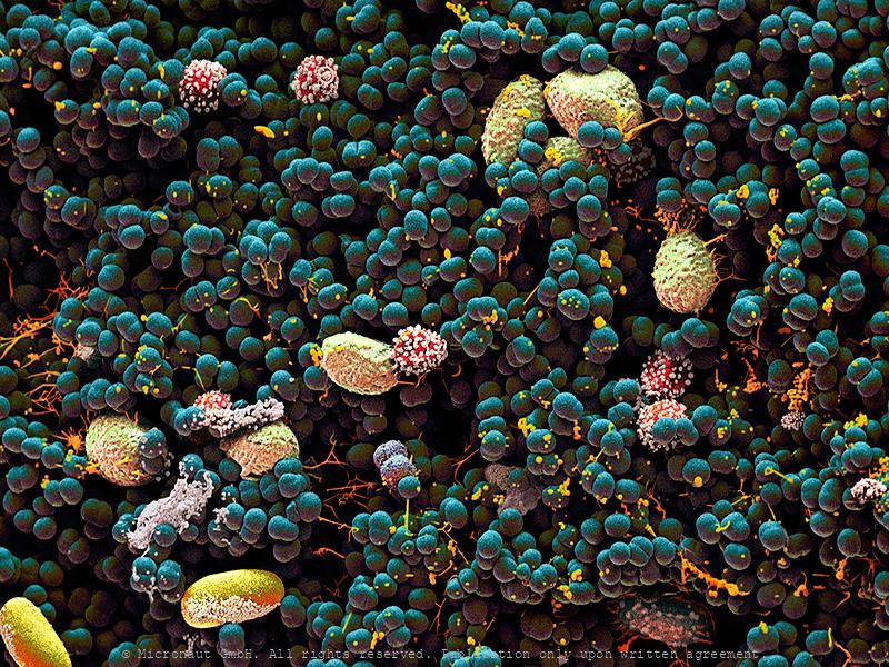
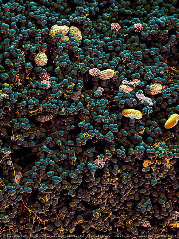
Belly Button Bacteria and Fungi Spores under the scanning electron microscope. Our body is home for lots of microbial species and interestingly, they have not been investigated until very recently. We imagine microbes as something bad, but that's not the case. In fact, most microbes on our body are either good for us or simply present, whether on your toe or in your nose. Right now on our bodies, bacteria are fighting other bacteria, fungi and even viruses. Among your skin’s true warrior clans are species of the genus Bacilllus such as Bacillus subtilis. Bacillus subtilis produces antibiotic compounds that can kill other bacteria and even foot fungi. Right now it may be doing this on your skin. We tend to think of the life on our skin as somehow stable and yet like any wild kingdom the individuals alive at any moment are the result of millions of independent wins and losses. Scientists have recently identified the Staphylococci, Corynebacteria, Actinobacteria, Clostridiales, and Bacilli as the most common groups of bacteria on our bodies. Some can be beautiful: - Species of Micrococcus (M. luteus) produce yellow colonies, others (M. roseus) red ones, at least when grown in agar. Micrococcus species are aerobes and can deal with extreme drought and long periods of starvation. This toughness predisposes them to a life spent on our desert-dry flesh. - The Clostridiales include bacteria like Anaerococcus and Clostridia. Despite some of them can be bad news, most are harmless or even beneficial. The Clostridiales are spindle-shaped and motile anaerobes. They may be growing in your belly button, or maybe inside your ear. - Like almost any bacteria in the wrong place, Staphylococcus species such as Staphylococcus epidermidis can become pathogens but when living on your skin, Staphylococcus are far more likely to be beneficial, working as our first line of defense against pathogen invasion.
Belly Button Bacteria
Belly Button Bacteria and Fungi Spores under the scanning electron microscope. Our body is home for lots of microbial species and interestingly, they have not been investigated until very recently. We imagine microbes as something bad, but that's not the case. In fact, most microbes on our body are either good for us or simply present, whether on your toe or in your nose. Right now on our bodies, bacteria are fighting other bacteria, fungi and even viruses. Among your skin’s true warrior clans are species of the genus Bacilllus such as Bacillus subtilis. Bacillus subtilis produces antibiotic compounds that can kill other bacteria and even foot fungi. Right now it may be doing this on your skin. We tend to think of the life on our skin as somehow stable and yet like any wild kingdom the individuals alive at any moment are the result of millions of independent wins and losses. Scientists have recently identified the Staphylococci, Corynebacteria, Actinobacteria, Clostridiales, and Bacilli as the most common groups of bacteria on our bodies. Some can be beautiful: - Species of Micrococcus (M. luteus) produce yellow colonies, others (M. roseus) red ones, at least when grown in agar. Micrococcus species are aerobes and can deal with extreme drought and long periods of starvation. This toughness predisposes them to a life spent on our desert-dry flesh. - The Clostridiales include bacteria like Anaerococcus and Clostridia. Despite some of them can be bad news, most are harmless or even beneficial. The Clostridiales are spindle-shaped and motile anaerobes. They may be growing in your belly button, or maybe inside your ear. - Like almost any bacteria in the wrong place, Staphylococcus species such as Staphylococcus epidermidis can become pathogens but when living on your skin, Staphylococcus are far more likely to be beneficial, working as our first line of defense against pathogen invasion.
Intestinal Bacteria
Human excrements with intestinal bacteria. The human gut teems with bacteria (streptococci, staphylococci, enterococci, enterobacteria, mycobacteria, spirochetes, mycoplasma, corynebacteria clostridia, or lactobacilli). Many of their species are still unknown. They help us digest food and absorb nutrients, and they play a part in protecting our intestinal walls. Gut bacteria may also help regulate weight and ward off autoimmune diseases. In adult humans, the intestine bacterial flore makes up about five percent of the body weight, forming a fragile ecosystem sensitive to stress, unhealthy food, alcohol or illness. Under unfavorable circumstances our health is directly affected and as a result we suffer from e.g. overweight, mood changes or prone to various diseases. In this sample, numerous intestinal bacteria can be detected. Additionally, a plant fiber has passed the intestinal tract unharmed and spans across the frame (center). On the right hand side (half-cut) the relatively large and spherical shaped cyste of a Giardia parasite (brown) is visible. Apart from this unusual parasitic supplement, the documented bacterial fauna looks fairly diverse and well-sorted…
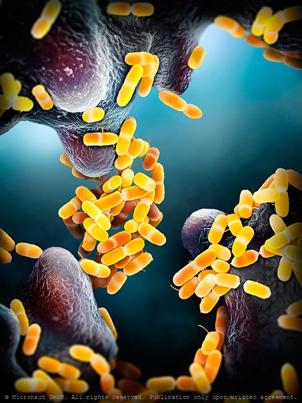
Army of Pathogens-Legionnaires' Disease (Legionella pneumophila)
When you finally return to work after the lockdown, coronavirus might not be the only illness you need to worry about contracting at the office. Office buildings once filled with employees emptied out in many cities and states as shelter-in-place orders were issued. These structures, normally in constant use, have been closed off and shut down, and health risks might be accumulating in unseen ways. Most buildings aren’t designed to be left alone for months. Therefore, public health authorities have issued warnings about the plumbing in these buildings, where water may have gone stagnant in the pipes or even in individual taps and toilets. As lockdowns are lifted, bacteria that build up internally may cause health problems for returning workers if the problem is not properly addressed by facilities managers. Employees and guests at hotels, gyms and other kinds of buildings may also be at risk. Some buildings may have remained closed for months due to the shutdowns, but the consequences of long-term water stagnation are relatively unknown according to researchers of the Perdue University. Typically, facilities managers reduce the risk of Legionella and other bacteria by pouring small amounts of disinfectant into a building’s water systems. But when the water is left stagnant for too long, the disinfectant disappears. “Even just after a weekend, disinfectant can be gone in some buildings and the water is vulnerable to contamination,” Dr. Whelton said. Even if only a small portion of buildings have problems, with so many reopening at once, the researchers fear there will be more outbreaks than usual. “Not every building will have issues but based on what we know, enough of them probably will,” Dr. Proctor said. (Ref. The New York Times, 27 Aug 2020).
Escherichia coli - Nr.5
Bacteria put the tang in yogurt and the sour in sourdough bread; bacteria help to break down dead organic matter; bacteria make up the base of the food web in many environments and they were among the earliest living organisms on earth. Escherichia coli bacteria are rod shaped and typically about 2µm long and 0.25-1µm in diameter. Some strains are motile and possess flagella. Living symbiotically inside the intestine of mammals, including humans, E. coli bacteria usually benefit their hosts by producing vitamin K2 and by preventing pathogenic bacteria to colonize the gut. Some strains produce a toxin that causes severe diarrhoea and can be fatal, especially in the very young or elderly. Furthermore, E. coli remains one of the most diverse bacterial species: Genome sequencing has revealed that only 20% of the genome is common to all strains (remember: the similarity of DNA between humans and chimpanzees is greater than 98%). Recently, scientists were able to engineer E. coli bacteria which can digest (or "upcycle") plastic waste. Thereby, Poly(ethylene terephthalate) (PET) is conversed into the small molecule vanillin, a flavour compound ubiquitous in the food and cosmetic industries, and an important bulk chemical. Currently a conversion rate of 79% is achieved. This represents the first biological upcycling process of plastic waste using an engineered microorganism.
The mutually beneficial relationship
Hand-colored Scanning-Electron-Micrograph of Rhizobium leguminosarium by Micronaut. Rhizobium is a genus of Gram-negative soil bacteria that fix nitrogen which forms an endosymbiotic nitrogen fixing association with roots of legumes. The bacteria colonize plant cells within root nodules where they convert atmospheric nitrogen into ammonia and then provide organic nitrogenous compounds such as glutamine or ureides to the plant. The plant in turn provides the bacteria with organic compounds made by photosynthesis. Beijerinck in the Netherlands was the first to isolate and cultivate a microorganism from the nodules of legumes in 1888, and named it Bacillus radicicola.
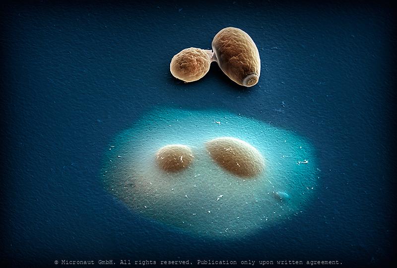
Encapsulated vs. normal yeast cells
Budding yeast (Saccharomyces cerevisiae); hand-colored scanning-electron-micrograph by Micronaut (Martin Oeggerli). Modification of cell surfaces with synthetic hydrogels is a promising approach for controlling cell behaviour. Here, Vanella and colleagues developed a genetically controlled system that enables cells to auto-encapsulate inside protective hydrogel capsules. The image shows an encapsulated budding yeast cell directly adjacent to a non-encapsulated one. This system links inducible gene expression to a synthetic capsule that protects target cells against lysis and mediates communication with the extracellular space through a permeable hydrogel.
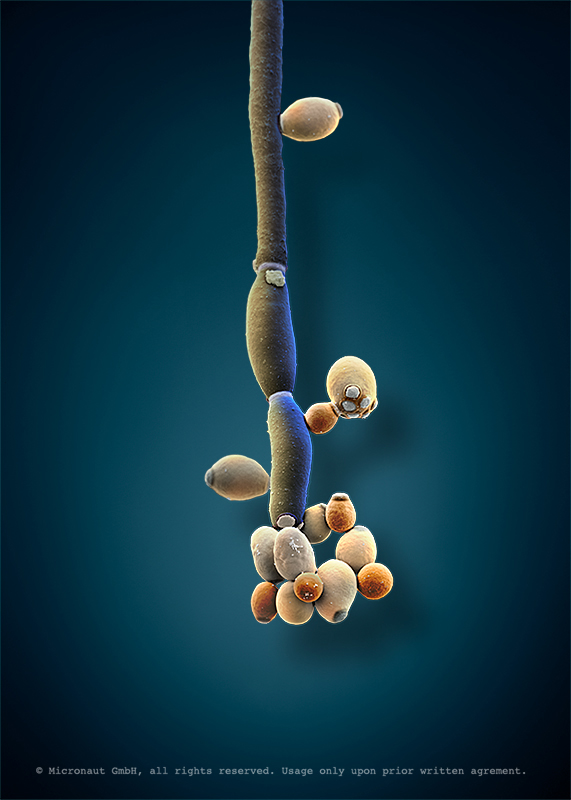
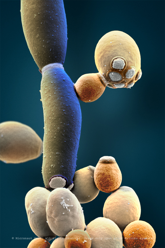
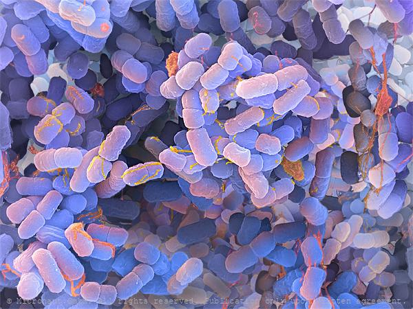
Face/Off - The interchangeable fungi (Candida albicans)
Candida albicans (laboratory culture), hand-colored SEM scan by Martin Oeggerli (Micronaut). Candida albicans is the most common member of the human gut flora and it can also survive outside the human body. In healthy human adults, it is a commensal microbe of the gastrointestinal tract and mouth in 40–60%. However, C. albicans can also become pathogenic in immunocompromised individuals, causing the human infection candidiasis, which results from an overgrowth of the fungus and can often be observed in e.g. in HIV-infected patients. Candidiasis is causing 2800-11200 deaths in the US yearly. Furthermore, C. albicans is the most common fungal species isolated from biofilms. It is also used as a model organism to study fungal pathogens. According to new studies, C. albicans can cross the blood–brain barrier in mice. Interestingly, C. albicans can grow as both, yeast (opaque and GUT form) and filamentous cells (pseudohyphal form): due to limited mobility, fungi have evolved mechanisms to adapt to sudden chemical and/or physical variation in their environment and scientists are using C. albicans to study eukaryotic responses to environmental changes by switching morphologies from yeast to hyphae growth.
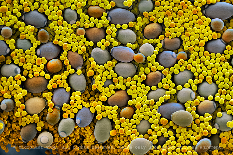
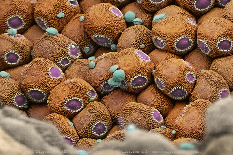
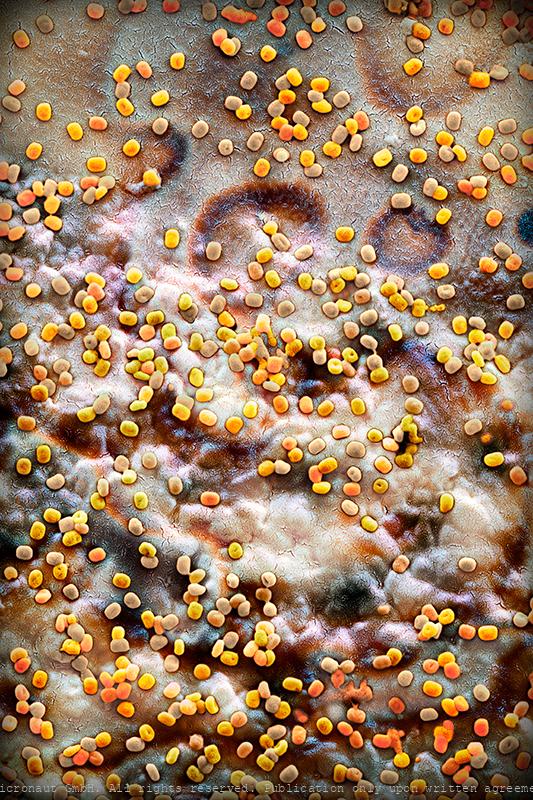
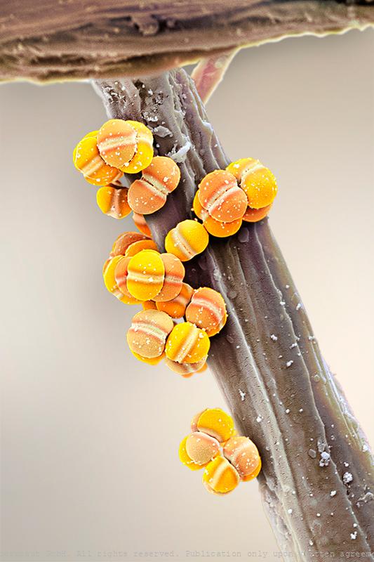
Blastoconidia of Candida albicans
Candida albicans (laboratory culture), hand-colored SEM scan by Martin Oeggerli (Micronaut). Candida albicans is the most common member of the human gut flora and it can also survive outside the human body. In healthy human adults, it is a commensal microbe of the gastrointestinal tract and mouth in 40–60%. It is a multi-morphologic creature with highly different structures: under certain environmental factors blastoconidia are produced. They are very small cells (the yellow cells) in this picture, produced asexually. Blastoconidia are able to divide and further spread within very short time. Despite it is a very common commensal species, C. albicans can also become pathogenic in immunocompromised individuals, causing the human infection candidiasis, which results from an overgrowth of the fungus and can often be observed in e.g. in HIV-infected patients. Candidiasis is causing 2800-11200 deaths in the US yearly. Furthermore, C. albicans is the most common fungal species isolated from biofilms. It is also used as a model organism to study fungal pathogens. According to new studies, C. albicans can cross the blood–brain barrier in mice. Interestingly, C. albicans can grow as both, yeast (opaque and GUT form) and filamentous cells (pseudohyphal form): due to limited mobility, fungi have evolved mechanisms to adapt to sudden chemical and/or physical variation in their environment and scientists are using C. albicans to study eukaryotic responses to environmental changes by switching morphologies from yeast to hyphae growth.
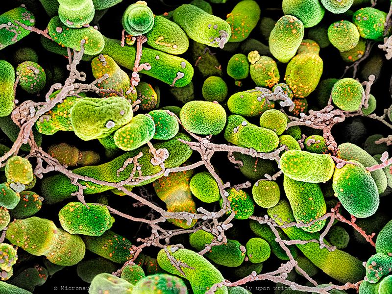
The Truffle (Tuber spp) microbiome
Truffles (i.e. the Périgord truffle Tuber melanosporum or the white truffle Tuber magnatum), Owing to their unique aromas, truffle fungi (Tuber spp.) are regarded as most precious food delicacies. Since many years, scientists try to decrypt aroma production in truffles. Over the last decade, Prof. Richard Splivallo has explored along with international colleagues the genomes of various truffle species as well as the specific contribution of bacteria in truffle aroma formation. These scientists could show that, based on the genomes, truffles seemed to possess most of the molecular machinery needed to produce many of their flavor compounds. Yet, Prof. Splivallo and colleagues could also show that bacteria colonizing truffle fruiting bodies were responsible for the production of key truffle volatiles. This illustrates that overall truffle aroma results from the intimate interaction of truffles with bacteria that are trapped inside of truffles. Manually colored scanning-electron-micrograph (SEM) by Martin Oeggerli, kindly supported by Richard Splivallo. The picture shows isolated bacteria from a truffle (Tuber melanosporum).
Waterbacteria (Caulobacter crescentus)
Water bacteria exist in two different forms: a mobile cell (called a “swarmer cell”) which is equipped with a hair-like flagellum to swim freely in the water and an immobile cell (called a “stalked cell”) which is equipped with an adhesive to attach the cell to the surface. Interestingly, both cells arise from asymmetrical cell division which is a complex and exceedingly rare process in nature and can e.g. lead to cancer. Waterbacteria allow scientists to study asymmetrical cell division in depth and to learn more about the process of cell division in general. Additionally, the stalked cell produces THE strongest glue in nature, for a safe anchorage to any surface and induces the formation of biofilms. It’s a nasty problem for the maritime industry, since biofilms increase drag and annually lead to additional costs and waste of fuel. Best scientific image 2008. -- Caulobacter teilt sich asymmetrisch in zwei Tochterzellen mit unterschiedlichem Entwicklungsprogramm. Die Bakterienart kann dadurch besser auf sich ändernde Umweltbedingungen reagieren. Forschungsversuche an Wasserbakterien dienen längerfristig dazu zelluläres Verhalten zu berechnen und einfache lebende Systeme zu konstruieren. Ausgezeichnet als Bestes Bild der Forschung 2008 (1. Rang Kategorie Faszination Forschung).
Waterbacteria
Waterbacteria (Caulobacter crescentus) attaching to a solid structure. Best scientific image 2008.
Anthrax (Bacillus anthracis)
Anthrax bacteria (Bacillus anthracis) is a Gram-positive endospore-forming rod more commonly called “anthrax”. In 2001 the organism was used as part of a terrorist attack on the USA, where a highly virulent strain of anthrax was mailed to senators killing 5 people. This has heightened the concern around anthrax as a biological weapon (Schwartz, 2009). B. anthracis is commonly found in soil where it can lie dormant, potentially for hundreds of years. Herbivores often disturb soil when grazing and can ingest or inhale the B. anthracis spores. The bacteria can be transmitted readily from close contact with animals, soil or through ingestion of contaminated meat (from an infected animal) (Schwartz, 2009). Occupations at high risk of infection include veterinary surgeons, livestock farmers, butchers and persons who handle hides or wool. Treatment and antibiotic resistance: Contrary to popular belief, antibiotics are very effective in controlling B. anthracis infections if used early enough. Antibiotics such as ciprofloxacin, doxycycline, erythromycin, vancomycin and penicillin are all useful for treatment of B. anthracis infections (Moayeri and Leppla, 2004). There is also a monoclonal antibody (Raxibacumab) that is currently showing promise in clinical trials , which can be used in the prophylaxic treatment of B. anthracis infection. B. anthracis has shown some resistance to antibiotics in laboratory testing and possesses antimicrobial enzymes such as b-lactamases (Bryskier, 2002). Prevention and control: Preventative measures include immunisation of humans and animals and post exposure antibiotic prophylaxis (Bryskier, 2002). The currently available vaccines include AVA (anthrax vaccine adsorbed), which is licensed in the USA and AVP (anthrax vaccine precipitated), which is licensed in the UK; these are used to create immunity to B. anthracis pre-exposure (Bryskier, 2002). Where people or animals have died from B. anthracis, great care must be taken to handle
Toxic Bacteria (Yersinia pestis)
Yersinia pestis (yellow bacteria with rod-like shape) is the causative agent of the systemic invasive infectious disease often referred to as the plague. Y. pestis are Gram-negative and can infect humans and other animals. Human Y. pestis infection takes three main forms: pneumonic, septicemic, and the notorious bubonic plagues which has had devastating effects on the human population throughout history.
Mycobacterium tuberculosis (red version)
Artistic coloration of a slowly growing colony of Mycobacterium tuberculosis which is a pathogenic bacterial species (family Mycobacteriaceae) and the causative agent of tuberculosis. M. tuberculosis is a small bacillus that can withstand weak disinfectants and can survive for weeks in a dry state. Its unusual cell wall is rich in lipids and responsible for this resistance. Cells are often seen wrapped together, due to the presence of fatty acids (e.g., mycolic acid) in the cell wall that stick together. This appearance is referred to as cording, like strands of cord that make up a rope. Dividing only every 15–20 hours, M. tuberculosis grows extremely slow compared with other bacteria, which tend to have division times measured in minutes rather than hours (E. coli: every 20 minutes). First discovered in 1882 by Robert Koch, M. tuberculosis is highly aerobic and requires high levels of oxygen. Primarily a pathogen of the mammalian respiratory system, it infects the lungs. Humans are the only known reservoirs of M. tuberculosis. When in the lungs, M. tuberculosis is taken up by alveolar macrophages, but they are unable to digest and eradicate the bacterium. Antibiotic resistance in M. tuberculosis typically occurs due to either the accumulation of mutations in the genes targeted by the antibiotic, or a change in titration of the drug. M. tuberculosis is considered to be multidrug-resistant (MDR TB) if it has developed drug resistance to both rifampicin and isoniazid, which are the most important antibiotics used in treatment.There are misconceptions that M. tuberculosis can be spread by shaking hands, making contact with toilet seats, sharing food or drink, sharing toothbrushes, or kissing. M. tuberculosis can only be spread through air droplets originating from a person either coughing, sneezing, speaking, or singing that has the disease. -- die Bakterien lagern sich z. T. zu zopfartigen Agglomeraten („Cord- Bildung“) zusammen
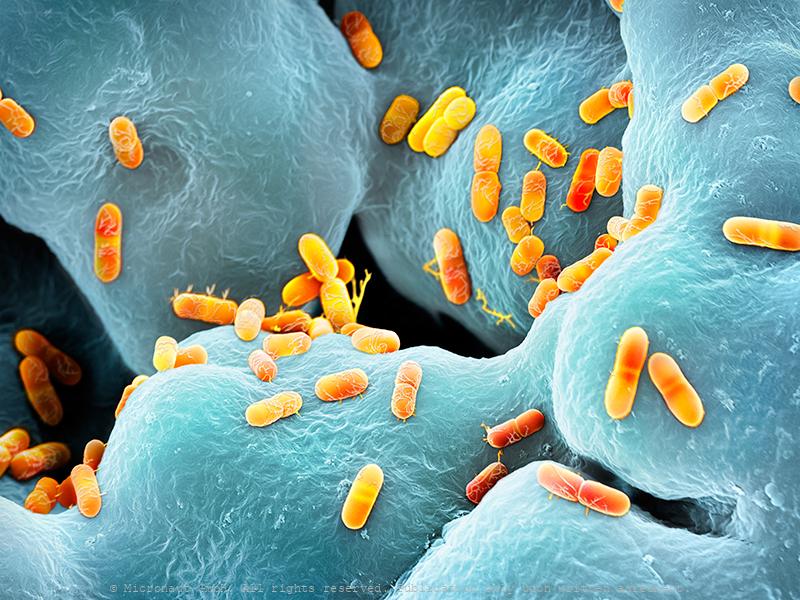
Escherichia coli bacteria, Nr.4
E. coli bacteria are living in the intestine of mammals, including humans.
The human gut flora - bacteria (Enterobacter cloacae)
Enterobacter cloacae bacteria, coloured scanning electron micrograph (SEM). Enterobacter cloacae is a clinically significant Gram-negative, facultatively-anaerobic, rod-shaped (bacillus) bacterium. Members of the genus Enterobacter exist in water, soil and the intestinal and urinary tracts of humans. In hospital patients with lowered immunity E. cloacae may be the causative agent of urinary tract and respiratory tract infections. In addition, it may also infect wounds on the skin, and quite often shows multiple resistancy to antibiotics.
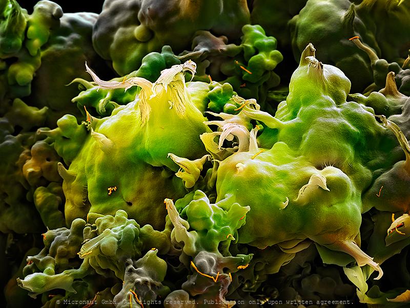
Stinky Foot Bacteria
What causes smelly feet? Sweat is released throughout the day to keep the skin moist and supple. Sweat itself is odorless, but creates a beneficial environment for certain bacteria species to grow and produce bad-smelling substances. The same species are naturally present on our skin as part of the human flora, but in rather low numbers. Some shoes and socks can inhibit evaporation and thus increase the amount of sweat you produce, thereby providing the perfect environment for 'stinky-feet-bacteria' to thrive. Since our feet have more sweat glands than on any other part of the body they typically start to smell bad first.
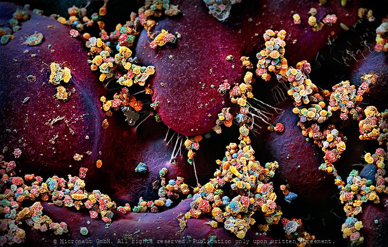
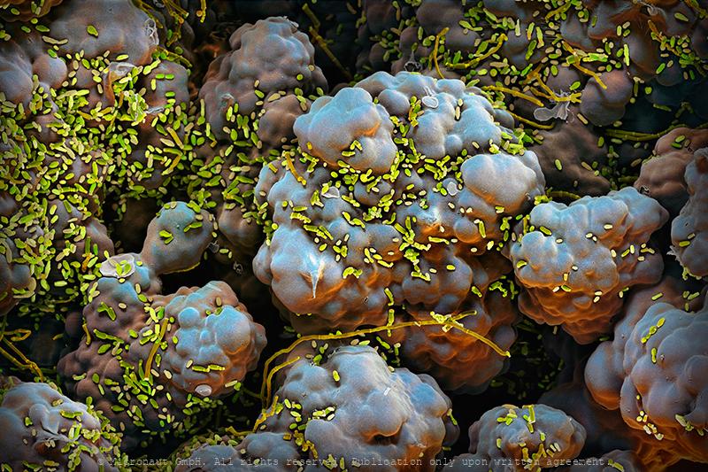
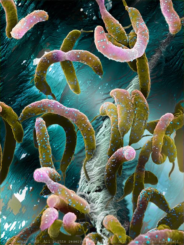
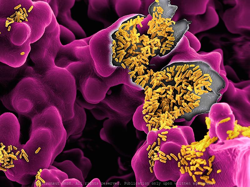
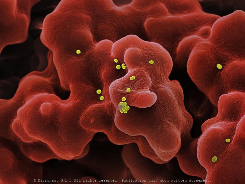
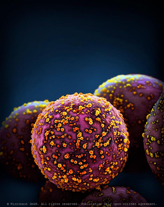
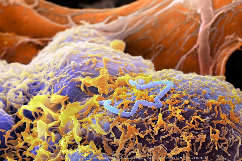
Stomach cells infection - Helicobacter pylori
In vitro cultured stomach cancer cells (red/brown), infected with Helicobacter pylori (yellow). Magenepithelzellen wurden in vitro gezüchtet und experimentell infiziert mit Helicobacter pylori; die Bakterien sind in gelb zu sehen, die Wirtszellen sind rot-braun. In vitro cultured stomach cancer cells (brown), infected with Helicobacter pylori (yellow). Helicobacter pylori, a common bacterium that lives in the stomach lining, increases the risk of stomach cancer and peptic ulcers. But over time H. pylori can reduce stomach acid and acid reflux, which may help fend off esophageal cancer. The microbe also appears to help protect us from allergies and asthma. Some scientists suspect that the dramatic increase in those conditions in the industrialized world could be related to the decreasing frequency of H. pylori in our stomachs, which is partly due to high doses of antibiotics in childhood.
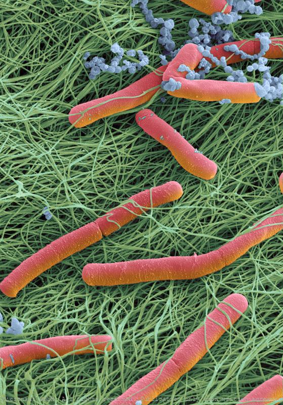
Globetrotters - Bacteria on a Toothpick (diverse species)
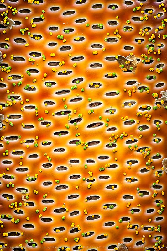
Microbiology explores the world of organisms too small to be seen without the aid of a microscope, such as bacteria, archaea, fungi, protozoans, microalgae, and viruses. Here you can see bacteria (mostly Streptococcus ssp.) found on a toothpick. The transportation system of plants includes invisibly small structures, called sieve-plates, that are involved in the transportation of sugars and other substances throughout the plant. A sieve-plate may contain hundreds of pores to absorb food particles and pass them on to the next sieve-element. Thereby sieve-plates regulate the size of particles which may travel within the plants vascular system. And in this case, they act as a beautifully shaped stage for the bacteria colony which was set-up in an in-vitro culture. This image has been produced using scanning electron microscopy (SEM) in combination with selectively assigning different colors to different bacteria species and structures. By creating a 3D-like photorealistic appearence, the artist aims to expose fascinating structures that are far too small to be seen with the naked human eye.
Dividing Streptococcus (beta-haem; remastered version)
dividing streptococcus sp. bacteria
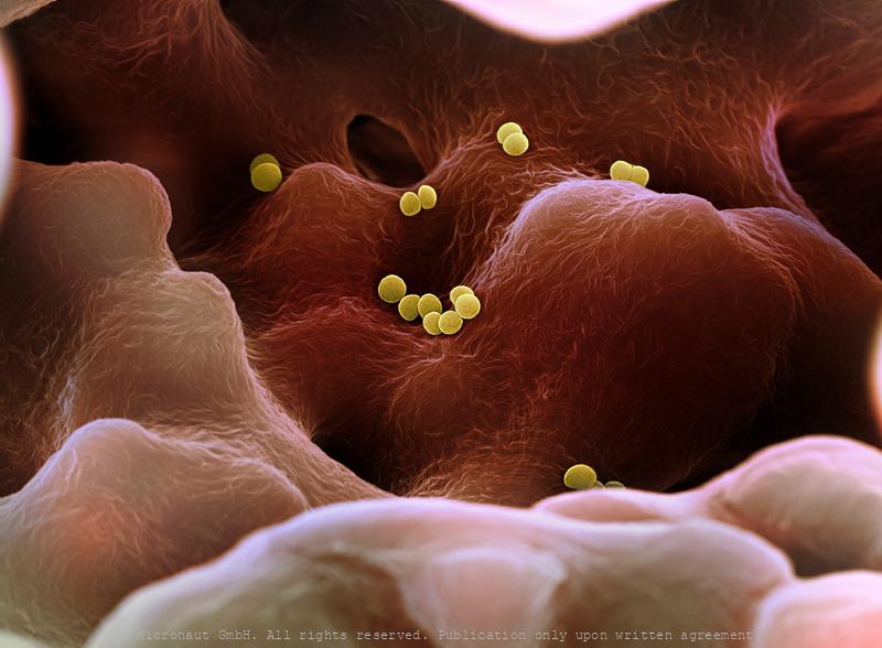
The Golden Staph (Staphylococcus aureus)
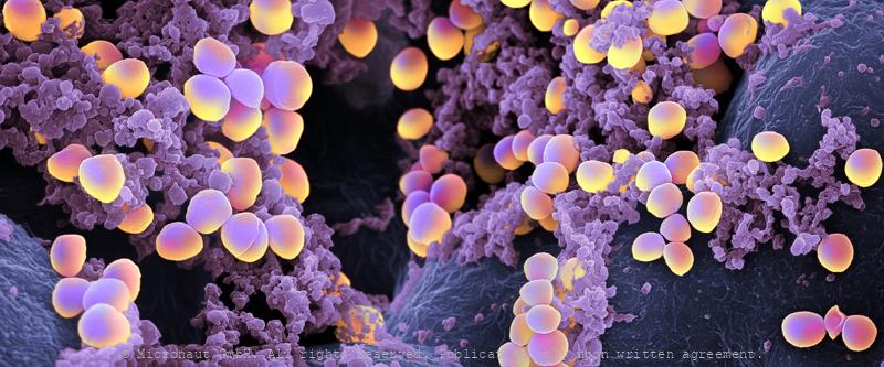
Staphylococcus aureus bacteria colony which has formed a biofilm. Staphs are among the most frequently used bac's in scientific research labs. 'Grape-shaped' S. aureus bacteria have started to develop a biofilm: after the bacteria adhere to the surface, they switch to the biofilm-mode of growth and produce a matrix of extracellular polymeric substance (EPS). The biofilm allows them to aggregate more easily and it also makes them increasingly resistant to antibiotics. Staphylococcus aureus Bakterienkolonie, welche damit begonnen haben einen Biofilm aufzubauen um besser aggregieren zu können und sich vor Antibiotikum zu schützen. S. aureus gehört zu den am häufigsten im Labor verwendeten Bakterienarten.
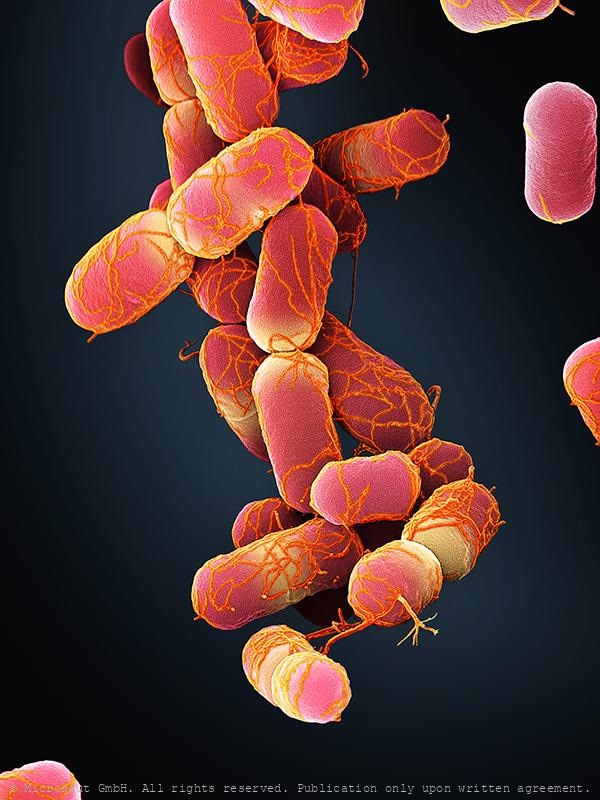
Escherichia coli bacteria, Nr.3 (red)
E. coli bacteria
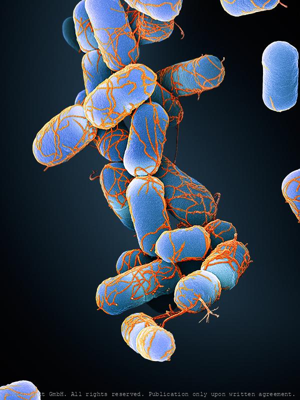
Escherichia coli bacteria, Nr.3 (blue)
E. coli bacteria
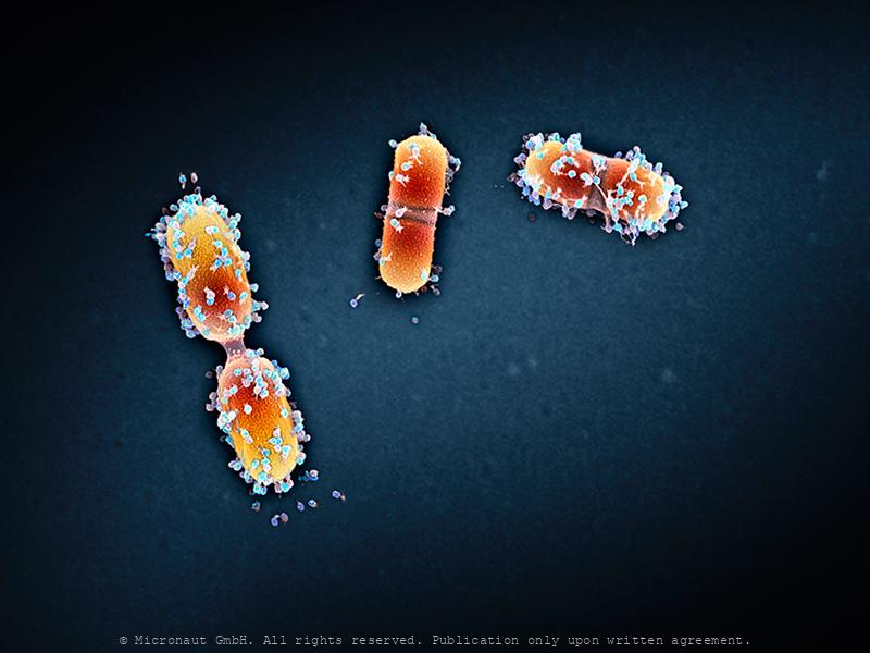
Viral infection - The Parasites of Parasites of Parasites... Nr.1
A bacteriophage is a virus that infects and replicates within a bacterium. It is composed of proteins that encapsulate a DNA or RNA genome. Bacteriophages ('phages') have relatively simple structures and their genomes encodes something between four and several hundred genes. To replicate, phages inject their genome into the cytoplasm of a bacteria. Bacteriophages are among the most common and diverse entities in the biosphere. They are widely distributed in locations populated by bacterial hosts, such as soil or the intestines of animals. One of the densest natural sources for phages and other viruses is sea water. This hand colored scanning-electron-micrograph shows bacteria which are heavily infected by bacteriophages.
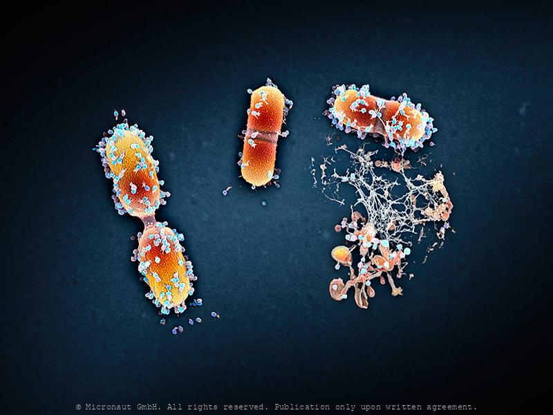
Viral infection - The Parasites of Parasites of Parasites... Nr.1
A bacteriophage is a virus that infects and replicates within a bacterium. It is composed of proteins that encapsulate a DNA or RNA genome. Bacteriophages ('phages') have relatively simple structures and their genomes encodes something between four and several hundred genes. To replicate, phages inject their genome into the cytoplasm of a bacteria. Bacteriophages are among the most common and diverse entities in the biosphere. They are widely distributed in locations populated by bacterial hosts, such as soil or the intestines of animals. One of the densest natural sources for phages and other viruses is sea water. This hand colored scanning-electron-micrograph shows bacteria which are heavily infected by bacteriophages. At the lower right hand side, a bacterial cell has been broken open (lysed) and destroyed. As soon as the cell is destroyed, the phage progeny can find new hosts to infect.
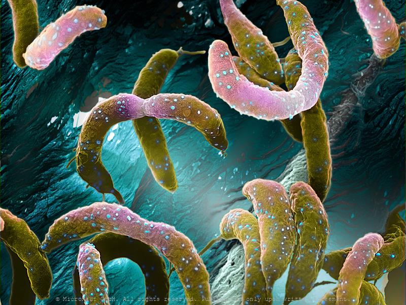
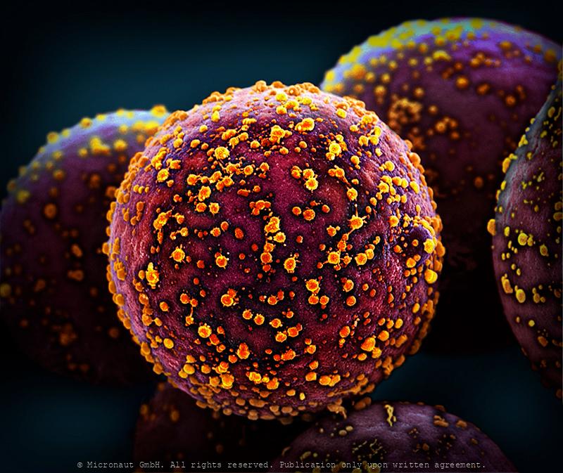
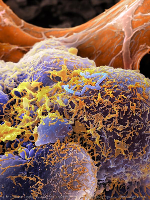
Bacteria (Helicobacter pylori)
In vitro cultured stomach cancer cells (red/brown), infected with Helicobacter pylori (yellow). Magenepithelzellen wurden in vitro gezüchtet und experimentell infiziert mit Helicobacter pylori; die Bakterien sind in gelb zu sehen, die Wirtszellen sind rot-braun. In vitro cultured stomach cancer cells (brown), infected with Helicobacter pylori (yellow). Helicobacter pylori, a common bacterium that lives in the stomach lining, increases the risk of stomach cancer and peptic ulcers. But over time H. pylori can reduce stomach acid and acid reflux, which may help fend off esophageal cancer. The microbe also appears to help protect us from allergies and asthma. Some scientists suspect that the dramatic increase in those conditions in the industrialized world could be related to the decreasing frequency of H. pylori in our stomachs, which is partly due to high doses of antibiotics in childhood.
Stinky Foot Bacteria
What causes smelly feet? Sweat is released throughout the day to keep the skin moist and supple. Sweat itself is odorless, but creates a beneficial environment for certain bacteria species to grow and produce bad-smelling substances. The same species are naturally present on our skin as part of the human flora, but in rather low numbers. Some shoes and socks can inhibit evaporation and thus increase the amount of sweat you produce, thereby providing the perfect environment for 'stinky-feet-bacteria' to thrive. Since our feet have more sweat glands than on any other part of the body they typically start to smell bad first.
The Human Microbiome, Nr.1
Artistic coloration of human excrements, that contain billions of intestinal bacteria, including Epulopiscium sp. - the world’s largest bacteria species! Artistic coloration of the microbiome present in a healthy human gut, containing billions of intestinal bacteria, including Epulopiscium sp. - the world’s largest bacteria species! The human gut teems with bacteria: streptococci, staphylococci, enterococci, enterobacteria, mycobacteria, spirochetes, mycoplasma, corynebacteria, clostridia, and lactobacilli. Many of their species are still unknown. They help us digest food and absorb nutrients, or play a role in protection of intestinal walls. Many gut bacteria further regulate weight and ward off autoimmune diseases. In adult humans, the intestinal flora is responsible for up to five percent of the body weight, forming a fragile ecosystem (the human microbiome) which is sensitive to: stress, unhealthy food, alcohol or illness. Under unfavorable circumstances our health is directly affected by bacteria that live inside of us, and as a result we may suffer from: overweight, mood changes or become prone to various diseases. In the central part of the image you can see the world’s largest bacteria - Epulopiscium sp. which spans between 200 – 700 µm, i.e. 350-times bigger than E. coli, or B. subtilis. Epulopiscium was first discovered in 1985 in the intestines of a surgeonfish, where it is only active during the day and believed to control the pH within the gut of its hosts. Because of its huge size, the giant exhibits many unusual characteristics, including a very special ‘rhomboid’ morphology. This image has been produced using scanning electron microscopy (SEM) in combination with selectively assigning different colors to different bacteria species and structures. By creating a 3D-like photorealistic appearence, the artist aims to expose the diversity of bacteria that can be found within human feces.
The Gut Microbiota Miracle - Microbiome of a Newborn (H. sapiens
The microbiome of a baby, 1 month after birth. Hand colored scanning electron micrograph by Martin Oeggerli (Micronaut). The Gut Microbiota Miracle: this picture shows the microbiome of a newborn one month after birth. The importance of our intestinal bacteria is already well known – but much less is known about its development and diversity from a newborn to the adult. It has long been assumed that human breast milk is sterile. But scientists have discovered complex and dynamic bacterial ecosystems in human breast milk which are distinct from other microbioal communities and may also colonize and widen the diversity of the infant gut probiotic bacterial community. According to the study, the cocktail of beneficial bacteria passed from mother to infant through breast milk changes significantly over time and could act like a daily booster for infant immunity and metabolism, with important implications for infant development and health. Bacteria found in breast milk include Actinobacteria, Bacillariaphyta, Bacteroidetes, Cyanobacteria, Deinococcus-thermus, Firmicutes, Fusobacteria, Patescibacteria, Plantae, and Proteobacteria.
E. coli bacteria Nr.1
Escherichia coli, highly enlarged. Biochemical manufacturers use various strains for biotransformations and biopharmaceutical production.
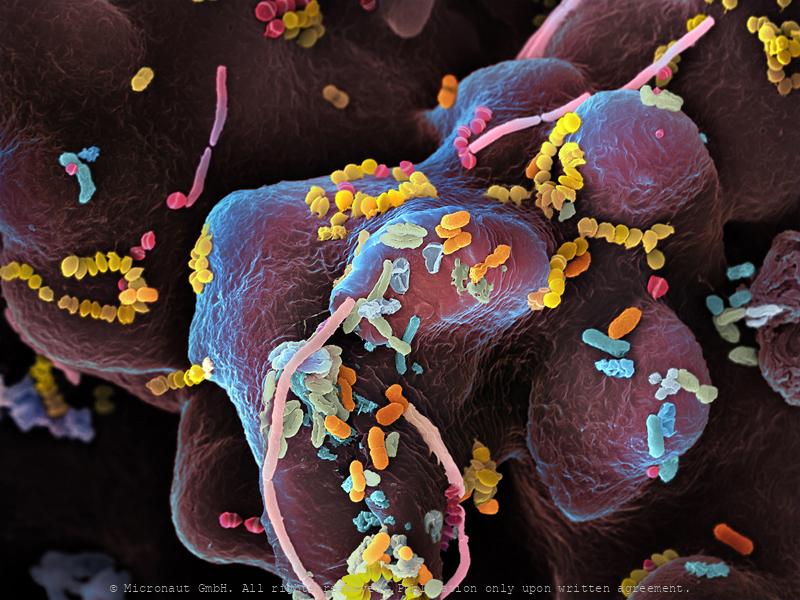
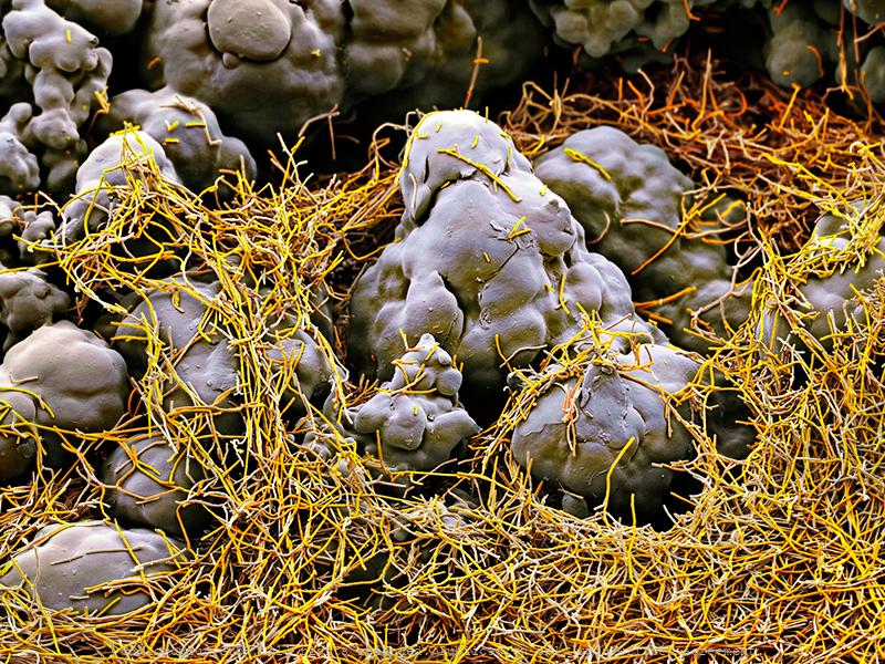
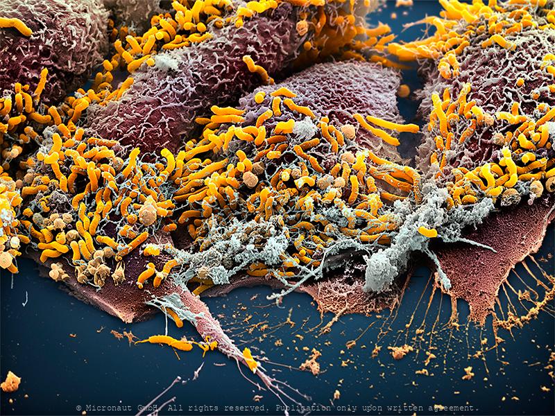
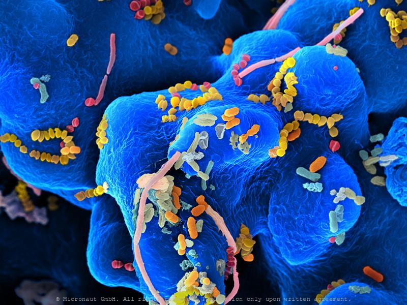
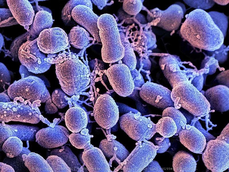
Oral bacteria
The human oral cavity contains a number of different habitats, including teeth, tongue, cheeks, hard and soft palates, which are colonized by bacteria. The oral microbiome is comprised of more than 600 identified species, and probably many more remain still unnamed. Some taking up residence on the mouth lining (red) within days after birth. Harmful species form biofilms, like the plaque that encourages tooth decay, or colonize the crevices between teeth and gums, causing periodontal disease. Oral probiotics designed to boost the population of species that outcompete pathogenic ones could help prevent or reverse dental disease.
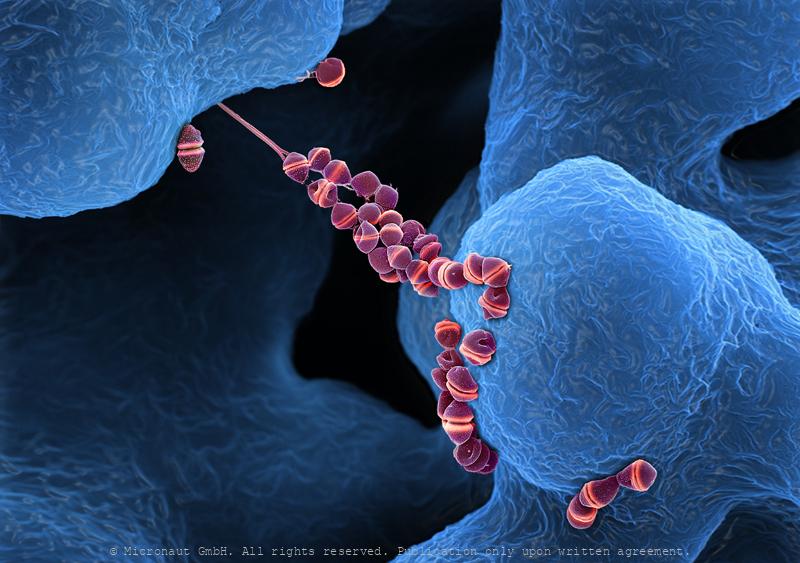
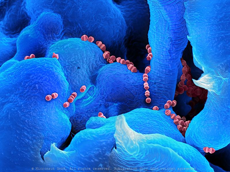
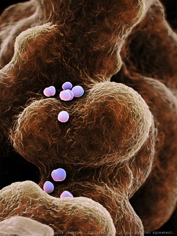
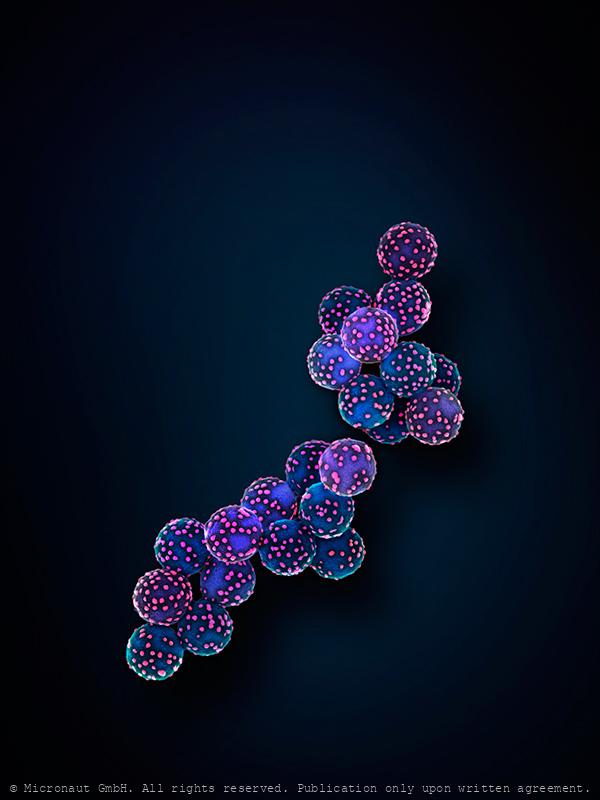
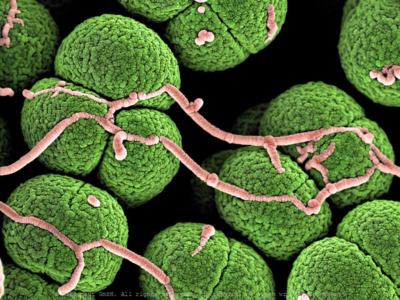
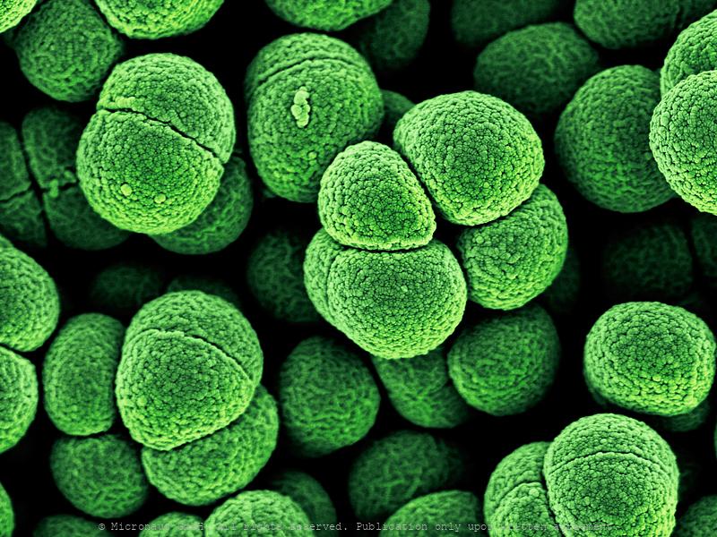
Streptococcus pneumoniae, Nr.2
Streptococcus pneumoniae are oval, gram-positive bacteria, that are often found in chains or pairs. They are responsble for many cases of meningitis, bacterial pneumonia and 'flesh-eating' bacterial infections. However, most species are non-pathogenic and are part of the normal commensal flora of the mouth, skin, intestine, and upper respiratory tract of humans. Interestingly, this species of bactera is able to grow very thin filaments, to climb directly from one peak to another.
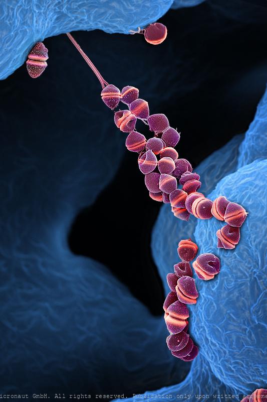
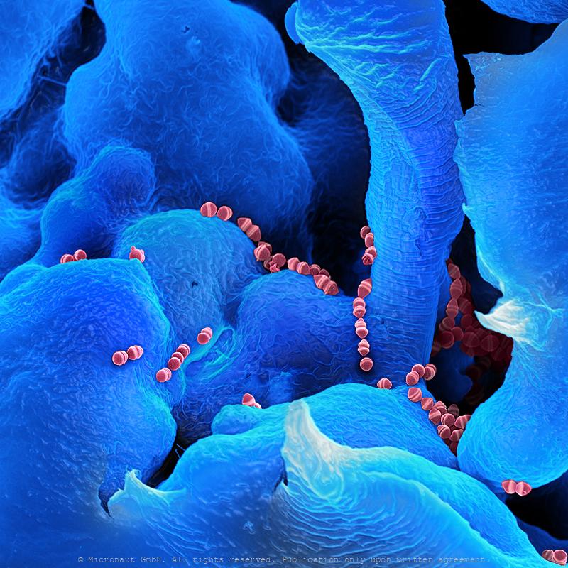
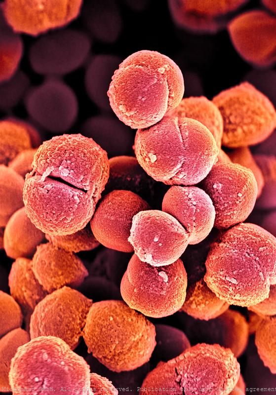
Streptococcus pneumoniae, Nr.1
Bacteria put the tang in yogurt and the sour in sourdough bread; bacteria help to break down dead organic matter; bacteria make up the base of the food web in many environments and they were among the earliest living organisms on earth. Streptococcus sp. bacteria are spherical shaped, typically 0.25-1µm in diameter. Bakterien (Staphylococcus pneumoniae)Bakterien sind unauffällige Erdenbewohner. 1676 wurden sie erstmals durch Antoni van Leeuwenhoek beschrieben. Während Bodenbakterien wichtige Nährstoffe für Pflanzen produzieren, leben Darmbakterien im Verdauungstrakt von Wiederkäuern, Pferden, oder Hasen. Dadurch werden Bakterien auch zur lebenswichtigen Nahrungsgrundlage für den Menschen. Handkoloriertes Raster-Elektronenmikroskopie Bild von Martin Oeggerli
Oral bacteria
The human oral cavity contains a number of different habitats, including teeth, tongue, cheeks, hard and soft palates, which are colonized by bacteria. The oral microbiome is comprised of more than 600 identified species, and probably many more remain still unnamed. Some taking up residence on the mouth lining (red) within days after birth. Harmful species form biofilms, like the plaque that encourages tooth decay, or colonize the crevices between teeth and gums, causing periodontal disease. Oral probiotics designed to boost the population of species that outcompete pathogenic ones could help prevent or reverse dental disease.
Staphylococcus infection, Nr.2
Staphylococcus infection, Nr.1
©-Micronaut-Bacteria-Staphylococcus-FHNW001c1_mini
Smallpox virus (Vaccinia sp.)
Conquered Killer – Smallpox ranks among the most devastating illnesses ever suffered by humankind. For centuries, it killed millions of people around the world. Thanks to global immunization programs, the deadly infectious disease was wiped out in 1979 following a successful vaccination program regarded as one of the greatest triumphs of modern medicine. Pockenviren sind die größten und bekanntesten Tierviren. Sie weisen Eigenschaften auf, die denen primitiver Zellen ähneln. Jedoch sind die Pockenviren, wie alle Viren, nicht in der Lage, Stoffwechsel zu betreiben, und sind daher in Bezug auf die Proteinbiosynthese vom Wirt abhängig. Diese Viren sind hinsichtlich DNA-Replikation ungewöhnlich: Eine mit dem Pockenvirus infizierte Zelle weist eine DNA-Synthese außerhalb des Kernes auf, was sonst nur in intrazellulären Organellen wie Mitochondrien (und bei Pflanzen in Chloroplasten) oder bei einer Infektion mit dem Hepatitis-B-Virus vorkommt. Bei den echten Pocken (Variola vera und Variola major) liegt die Sterblichkeit bei 10-90%. Die Übertragung erfolgt über Tröpfcheninfektion, zB bei Husten. Beim Krankheitsverlauf kommt es zu auffälligen Hautveränderungen (Pustel, Eiterbläschen). Für die Impfung werden Kuhpocken (Vaccinia Viren) verwendet. Diese sind im Bild zu sehen.
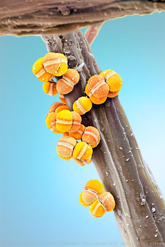
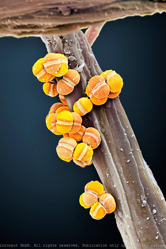
Rust fungus spores
hese fungal spores inside the fungal fruitbody of a Rust fungus (Urediniomycetes sp.) wait to be dispersed by the wind. Image was awarded 'Best Scientific Cover Image 2008'.
Fungal attack
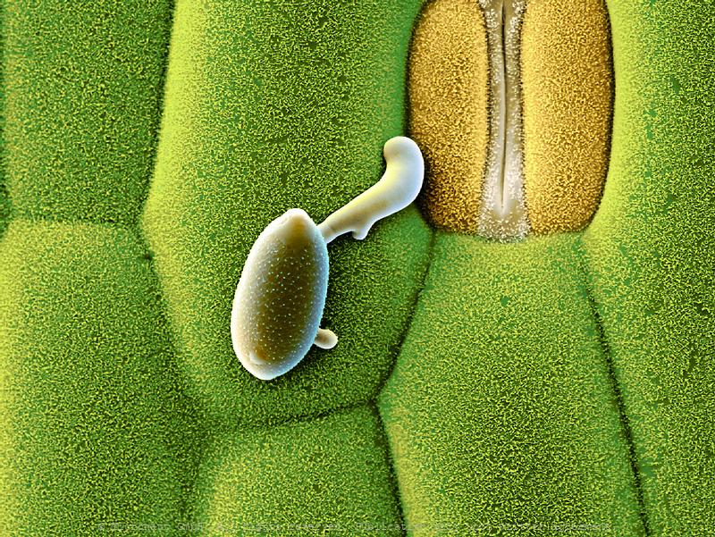
Fungi spore sitting and waiting in front of a plant pore (Wheat)
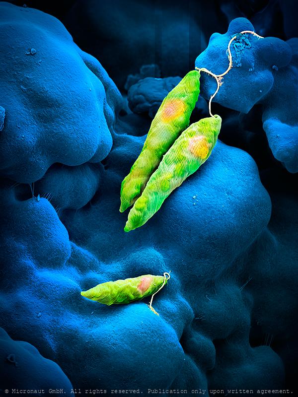
Flagellate (Euglena sp.)
Euglena is a genus of unicellular flagellate protists. It is the best known and most widely studied member of the phylum Euglenozoa, a diverse group containing some 44 genera and at least 800 species. Most species of Euglena have photosynthesizing chloroplasts within the body of the cell, which enable them to feed by autotrophy, like plants. However, they can also take nourishment heterotrophically, like animals. Euglena possess a red eyespot, an organelle composed of carotenoid pigment granules. The red spot itself is not thought to be photosensitive. Rather, it filters the sunlight that falls on a light-detecting structure at the base of the flagellum (a swelling, known as the paraflagellar body), allowing only certain wavelengths of light to reach it. As the cell rotates with respect to the light source, the eyespot partially blocks the source, permitting the Euglena to find the light and move toward it (a process known as phototaxis). Euglena lacks a cell wall. Instead, it has a pellicle made up of a protein layer supported by a substructure of microtubules, arranged in strips spiraling around the cell. The action of these pellicle strips sliding over one another gives Euglena its exceptional flexibility and contractility.
Rhizobium sp. Nr.1
Rhizobium sp. highly enlarged. This bacteria species is used for the biosynthesis fo L-carnitine
Rod-shaped bacteria
Virus Imprinted Nanomaterial Particles
Artificial virus recognition nanomaterials- Scanning electron micrograph of virus imprinted particles - VIPs - created via a surface initiated bottom-up synthetic approach using viruses as templates. The blue particles (approx. 400 nm) represent the VIPs while the "pink" dots represent the trapped virus.
Beads with Viruses
Beads with viruses
Beads with Viruses
Beads with viruses
Smallpox virus (Vaccinia sp.)
Conquered Killer – Smallpox ranks among the most devastating illnesses ever suffered by humankind. For centuries, it killed millions of people around the world. Thanks to global immunization programs, the deadly infectious disease was wiped out in 1979 following a successful vaccination program regarded as one of the greatest triumphs of modern medicine. Pockenviren sind die größten und bekanntesten Tierviren. Sie weisen Eigenschaften auf, die denen primitiver Zellen ähneln. Jedoch sind die Pockenviren, wie alle Viren, nicht in der Lage, Stoffwechsel zu betreiben, und sind daher in Bezug auf die Proteinbiosynthese vom Wirt abhängig. Diese Viren sind hinsichtlich DNA-Replikation ungewöhnlich: Eine mit dem Pockenvirus infizierte Zelle weist eine DNA-Synthese außerhalb des Kernes auf, was sonst nur in intrazellulären Organellen wie Mitochondrien (und bei Pflanzen in Chloroplasten) oder bei einer Infektion mit dem Hepatitis-B-Virus vorkommt. Bei den echten Pocken (Variola vera und Variola major) liegt die Sterblichkeit bei 10-90%. Die Übertragung erfolgt über Tröpfcheninfektion, zB bei Husten. Beim Krankheitsverlauf kommt es zu auffälligen Hautveränderungen (Pustel, Eiterbläschen). Für die Impfung werden Kuhpocken (Vaccinia Viren) verwendet. Diese sind im Bild zu sehen.
Stinky Foot Bacteria
What causes smelly feet? Sweat is released throughout the day to keep the skin moist and supple. Sweat itself is odorless, but creates a beneficial environment for certain bacteria species to grow and produce bad-smelling substances. The same species are naturally present on our skin as part of the human flora, but in rather low numbers. Some shoes and socks can inhibit evaporation and thus increase the amount of sweat you produce, thereby providing the perfect environment for 'stinky-feet-bacteria' to thrive. Since our feet have more sweat glands than on any other part of the body they typically start to smell bad first.
Coronavirus Structures - CoV2
Coronavirus structures. 9 original TEM image scans, hand-colored by Martin Oeggerli.
Doxycycline - Antibiotics
Antibiotics (Doxycycline)
Bacteria
Bacs
E. coli bacteria Nr.1
Escherichia coli, highly enlarged. Biochemical manufacturers use various strains for biotransformations and biopharmaceutical production.
Staphylococcus bacteria
Staphylococcus bacteria
S. ureae bacteria
Sporosacina sp. highly enlarged. These bacteria are used in scientific training, biosynthesis and biocatalysis.
Gluconobacter
Gluconobacter oxydans are found in flowers, fruits, garden soil, alcoholic beverages, cider and soda. They may be non-pathogenic to humans and animals, but cause apples and pears to rot and turn shades of brown. These microbes have limited metabolic abilities; partially oxidoze carbohydrates and alcohol through oxidative fermentation. Used to synthesis vitamin C, D-gluconis acid and ketogluconic acids. Can be used to convert glycerol to dihydroxyacetone since they thrive in environments wiht high consentration of sugar. Their biotechnological significance comes from the ability to incompletely oxidoze carbon substrates.
©-Micronaut-Bacteria-Lysteria-FHNW004a1_mini2_crop
©-Micronaut-Bacteria-Lysteria-FHNW004a1_mini2