Cell Wars
A century ago the first set of rules for hobby war games was established by HG Wells. Some hardcore gamers are still playing according to these, while millions more are playing games that have evolved out of the beginnings. However, there is a much larger battleground in everyone of us: the human body contains billions of participants (the cells) which are constantly arising & dying, interacting, communicating, evolving, or fighting against pathogens, etcetera. In fact, the list of what is happening in our body is endless – every second 50 million cells are dying inside of us and need to be replaced to let us continue to thrive and exist. The human body is a fascinating place to explore and would be the largest and most complex game by far, if we could program it – but it’s simply beyond our imagination! In part it’s because these processes are well hidden, taking place in a very small dimension and are technically extremely difficult and time consuming to analyze. Also, what happens cannot always be visualized, because it’s either too fast, or too slow, or we just lack the technology to measure and understand it. Here, we are using the scanning-electron-microscope to throw a glance into this facinating wold.
«These images are really outstanding. They raise all sorts of research questions I had never even considered before.»
Rob R. Dunn, PhD
Prof. of Applied Ecology and Book author
Micronaut images are rights-managed. If you want to get a quote, please contact us, providing the following information: (1) image name, (2) specific use, (3) industry, (4) distribution area, (5) format, (6) circulation or print run, and (7) duration.
Please note that we cannot answer incomplete requests. Thank you.
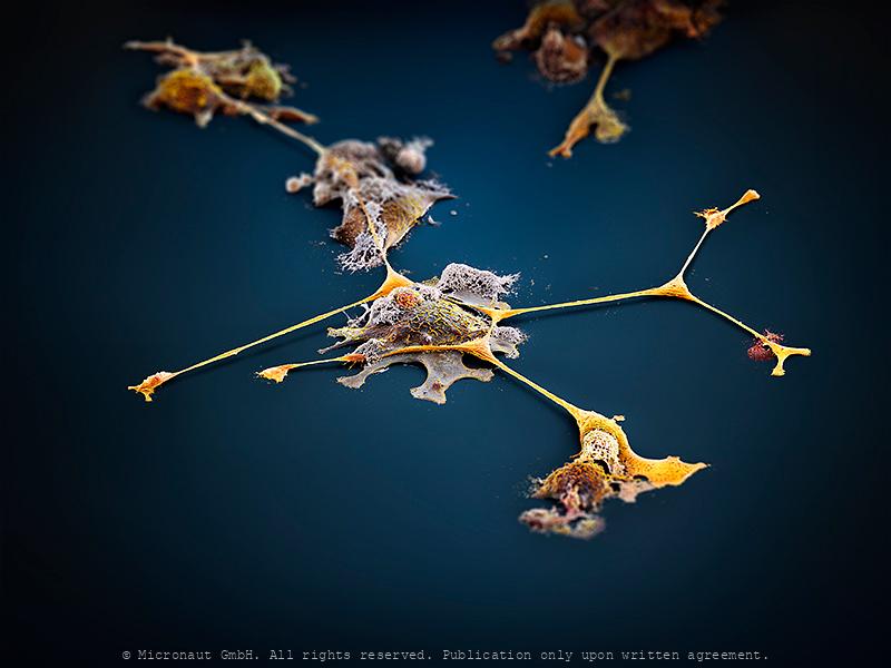
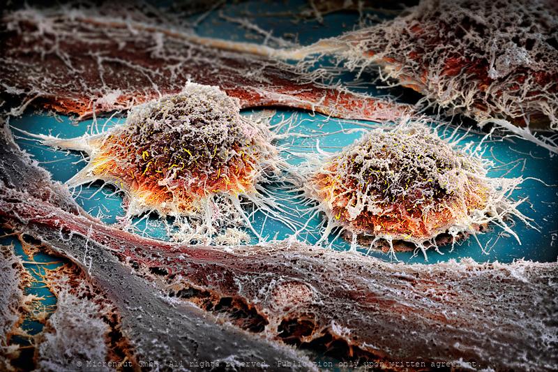
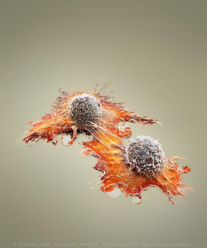
Cellular Islands (H. sapiens)
Tumors contain a vastly complicated cellular network that relies on local communication to execute malignant programs. The molecular cues that are involved in cell-cell adhesion orchestrate large-scale tumor behaviors such as proliferation and invasion. We have recently begun to understand that many tumors contain a high degree of cellular heterogeneity and are organized in a cellular hierarchy, with a cancer stem cell (CSC) population identified at the apex in multiple cancer types.
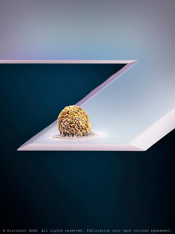
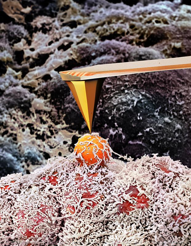
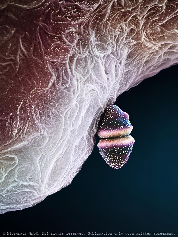
Weight-in of a cell (H. sapiens)
human Cell on an AFM cantilever. Atomic force microscopy (AFM) is a powerful, multifunctional imaging platform that allows biological samples, from single molecules to living cells, to be visualized and manipulated. The combination of AFM-based imaging and spectroscopy allows 3D manipulation with molecular precision. In this picture, we measure the weight of a cell with with AFM (cantilever). It is anticipated that in the next decade AFM-related techniques will have a profound influence on the way researchers view, characterize and solve fundamental challenges that cannot be addressed with other techniques.
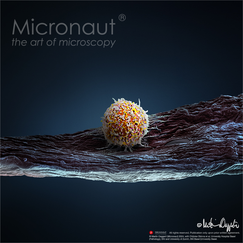
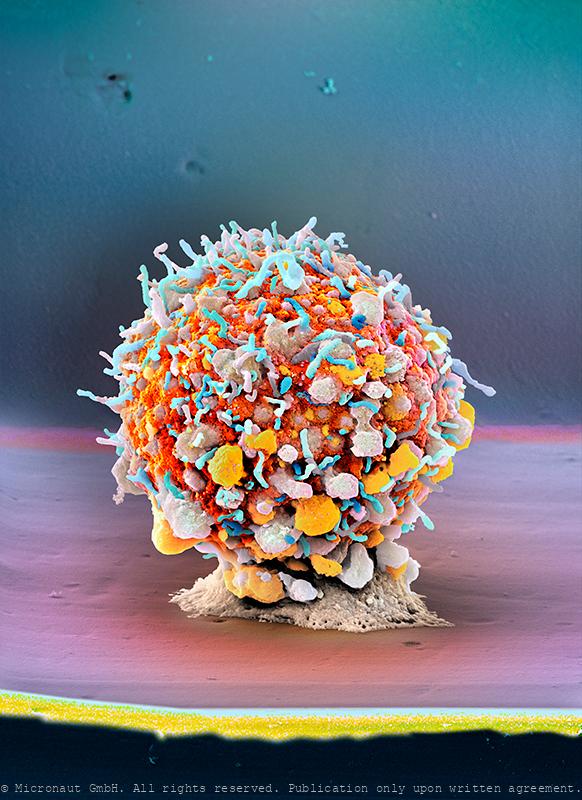
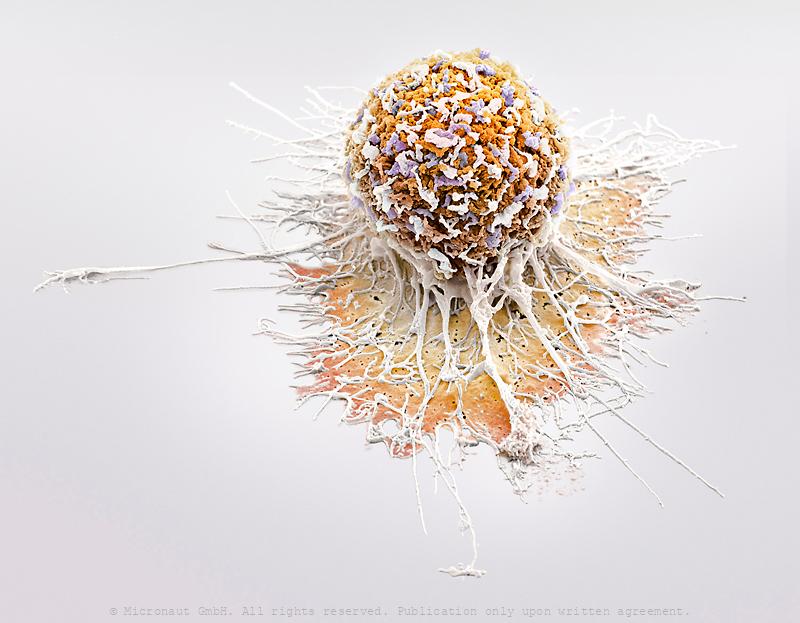
human T cell - engineered immune cell to fight cancer
Cellular Immunotherapy: this picture shows a human T cell from a immunocompetent humanized tumour model. Cancer immunotherapy with patients‘ own immune cells engineered to express chimeric antigen receptors (CARs) is a revolutionizing new treatment modality in hematologic oncology. CAR T cell therapy has achieved complete remission rates of 50 - 90% in patients with chemotherapy refractory or relapsed hematologic malignancies like diffuse large B cell lymphoma (DLBCL). By comparison, in DLBCL for example, the complete response rate to conventional therapies in patients with refractory or relapsed disease is below 10%. Still, up to one third of patients treated with CAR T cells relapse within a year after an initial complete response, there is a high burden of therapy associated toxicities and non-hematologic cancers have thus far not been amenable to redirected cellular immunotherapy. Apart from T cells, natural killer (NK) cells are a second cytotoxic lymphocytic lineage that have not been sufficiently explored clinically in the context of CAR therapy. Indeed, NK cells might be a promising alternative to T cells due to an inherent anti-cancer activity and a favorable toxicity profile. With a longstanding interest in human NK cells, our research includes engineering these cells as vehicles for redirected cellular immunotherapy. As a preclinical cancer model, we are using human immune system (HIS) mice carrying EBV-induced lymphomas. This allows the exploration of an autologous human cancer-immune system interface and its manipulation. We are specifically interested in engineering cytotoxic cells beyond redirection to enhance anti-cancer activity of the infused cell product. Another interest in the lab is functional analysis of genes important for human NK cell biology in vivo using HIS mice via genome editing approaches in the context of viral infection and homeostasis. These studies are designed to contribute to novel insights and improvements in the therapeuti
Quasi una Fantasia - The Dancing Hamster Cell (2/3)
Tracing back a hamster’s journey from the deserts of China to the research labs of Harvard in the mid-50s and on to becoming the golden standard for the bio-manufacturing industry and eventually - a work of art. Since they were first isolated in the late 1950s, Chinese hamster ovary (CHO) cells travel on an amazing journey. Because they are so easy to grow much was already known about their genetics and growth characteristics, CHO cells moved into the spotlight of the growing bio-pharmaceutical industry, which is currently worth more than $ 125 billion per year in sales at high profit margins. The step that catapulted CHO cells into prominence was the FDA approval for bio-therapeutic protein production - combined with an ability to grow in suspension, which greatly facilitates cell culturing at large scale. Meanwhile, CHO have become the golden standard for the production of therapeutic proteins, including humanized antibodies, and continue an unbelievable voyage all around the world. This hand-colored scanning-electron-micrograph (SEM) shows a chinese hamster (Cricetulus griseus) ovary cell on a phonograph record of Ludwig van Beethoven’s Moonlight Sonata (Sonata No 14 in C). The phonograph record was the primary medium used to store and reproduce music from the late 1880s. Records were later mainly made of Polyvinyl and retained the largest market share for about 100 years. Even after the introduction of the compact disc, and other new digital media formats, the Vinyls continue to be used and listened by DJs and a growing niche market of audiophiles… and they even serve as a perfect stage to present the worlds most famous cell - as work of art.
Quasi una Fantasia - The Dancing Hamster Cell (3/3)
Tracing back a hamster’s journey from the deserts of China to the research labs of Harvard in the mid-50s and on to becoming the golden standard for the bio-manufacturing industry and eventually - a work of art. Since they were first isolated in the late 1950s, Chinese hamster ovary (CHO) cells travel on an amazing journey. Because they are so easy to grow much was already known about their genetics and growth characteristics, CHO cells moved into the spotlight of the growing bio-pharmaceutical industry, which is currently worth more than $ 125 billion per year in sales at high profit margins. The step that catapulted CHO cells into prominence was the FDA approval for bio-therapeutic protein production - combined with an ability to grow in suspension, which greatly facilitates cell culturing at large scale. Meanwhile, CHO have become the golden standard for the production of therapeutic proteins, including humanized antibodies, and continue an unbelievable voyage all around the world. This hand-colored scanning-electron-micrograph (SEM) shows a chinese hamster (Cricetulus griseus) ovary cell on a phonograph record of Ludwig van Beethoven’s Moonlight Sonata (Sonata No 14 in C). The phonograph record was the primary medium used to store and reproduce music from the late 1880s. Records were later mainly made of Polyvinyl and retained the largest market share for about 100 years. Even after the introduction of the compact disc, and other new digital media formats, the Vinyls continue to be used and listened by DJs and a growing niche market of audiophiles… and they even serve as a perfect stage to present the worlds most famous cell - as work of art.
Quasi una Fantasia - The Dancing Hamster Cell (1/3)
Tracing back a hamster’s journey from the deserts of China to the research labs of Harvard in the mid-50s and on to becoming the golden standard for the bio-manufacturing industry and eventually - a work of art. Since they were first isolated in the late 1950s, Chinese hamster ovary (CHO) cells travel on an amazing journey. Because they are so easy to grow much was already known about their genetics and growth characteristics, CHO cells moved into the spotlight of the growing bio-pharmaceutical industry, which is currently worth more than $ 125 billion per year in sales at high profit margins. The step that catapulted CHO cells into prominence was the FDA approval for bio-therapeutic protein production - combined with an ability to grow in suspension, which greatly facilitates cell culturing at large scale. Meanwhile, CHO have become the golden standard for the production of therapeutic proteins, including humanized antibodies, and continue an unbelievable voyage all around the world. This hand-colored scanning-electron-micrograph (SEM) shows a chinese hamster (Cricetulus griseus) ovary cell on a phonograph record of Ludwig van Beethoven’s Moonlight Sonata (Sonata No 14 in C). The phonograph record was the primary medium used to store and reproduce music from the late 1880s. Records were later mainly made of Polyvinyl and retained the largest market share for about 100 years. Even after the introduction of the compact disc, and other new digital media formats, the Vinyls continue to be used and listened by DJs and a growing niche market of audiophiles… and they even serve as a perfect stage to present the worlds most famous cell - as work of art.
Dendritic Cells (H. sapiens)
Artistic coloration of a human dendritic cell, illustrating the characteristic ‘star-like’ shape and indicating the central role dendritic cells play for the immune system. Dendritic cells are antigen-presenting cells of the mammalian immune system. Their main function is to process antigen material and present it on the cell surface to the T cells of the immune system. Once activated, dendritic cells can migrate to lymph nodes where they interact with T cells and B cells to initiate an adaptive immune response. At certain development stages they grow branched projections, the dendrites (or "veils") that give the cell its name. Dendritic cells are also able to develop sheet-like processes that fold back onto the membrane surface. When exposed to e.g. HIV, these sheets entrap viruses in the vicinity and focus them to contact zones with T-cells targeted for infection. The dendritic cell was first described by Paul Langerhans (hence "Langerhans cell") in the late nineteenth century. In 1973 Ralph M. Steinman and Zanvil A. Cohn established the term "dendritic cell". For discovering the central role of dendritic cells in the adaptive immune response, Steinman was awarded the Nobel Prize in Physiology or Medicine in 2011.This image was produced using scanning electron microscopy in combination with selectively assigning different colors to different structures. By creating a 3D-like photorealistic appearance, the artist aims to expose the complex structure and cellular interaction of an activated dendritic cell.
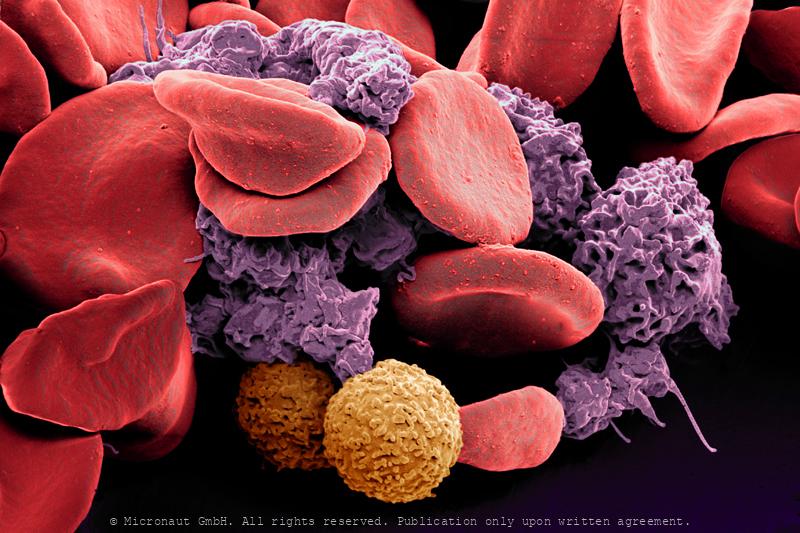
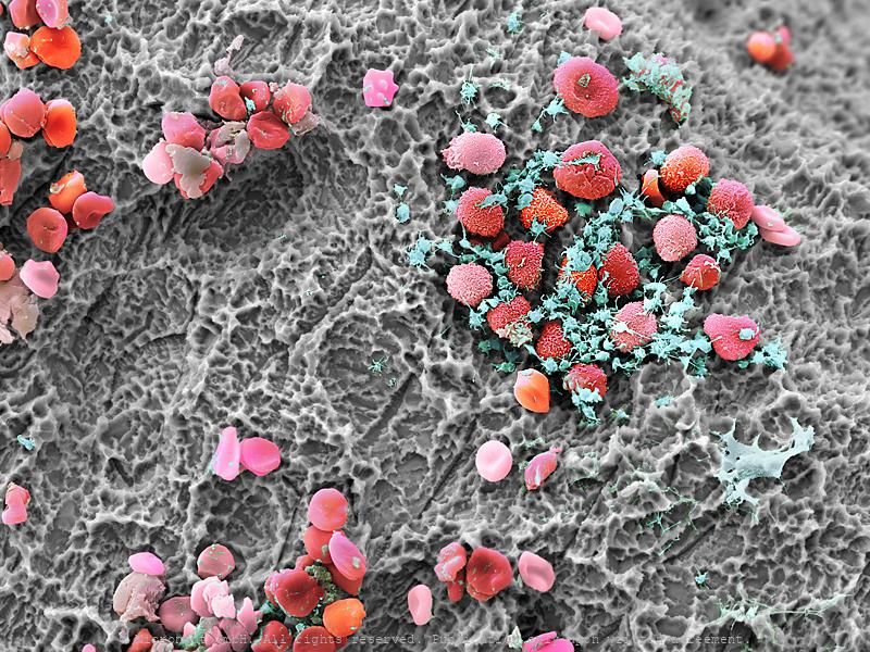
Blood cells
Blood cells
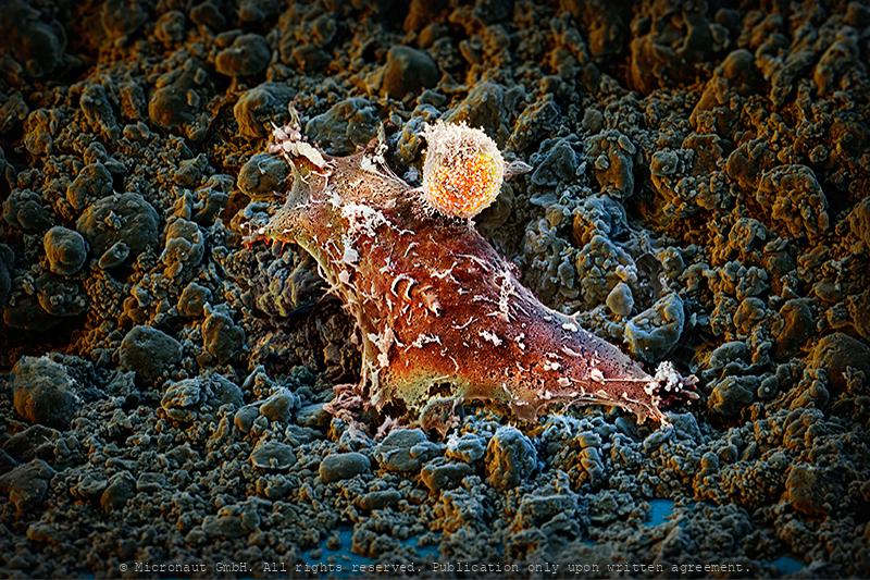
Cell Wars
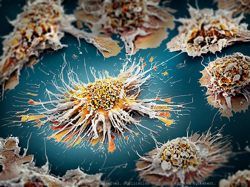
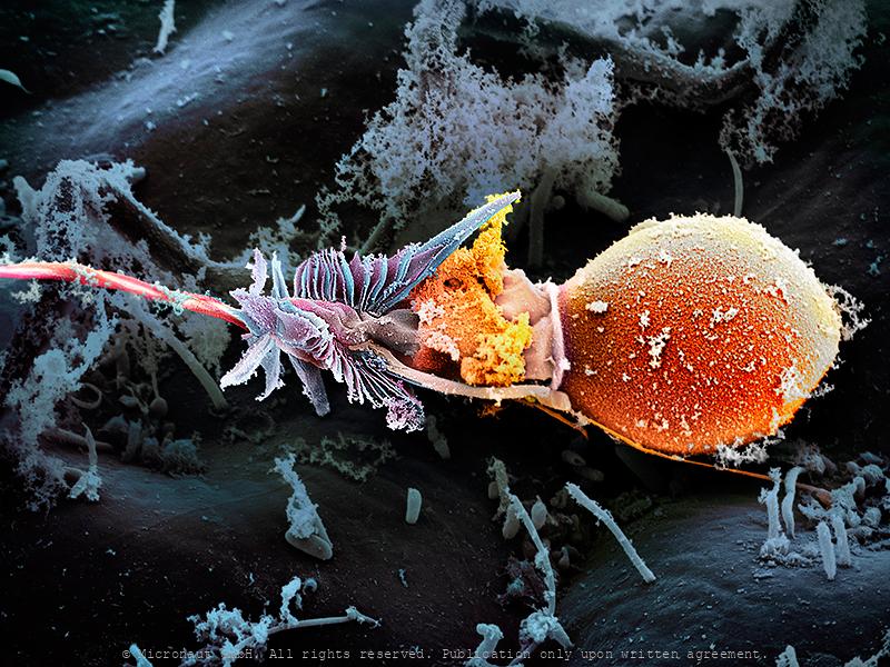
The micrograph shows a perforin-positive Interleukin-2 activated natural killer (NK-) lymphocyte in action. The missile like NK killer attacking a brain tumor (glioblastoma) cell was fixed exactly when it inserted its deadly enzymes into its target. Membrane protrusions of the killer resemble plant root formations; the upper right of the glioblastoma target shows a ruffled surface where a killer had been previously attached. Some membrane remnants remain. This structure-function relationship between a highly active killer and its target is totally new to the scientific and non-scientific community. It further demonstrates, that also healthy individuals bear immune surveillance elements against deadly brain tumor cells. Die Killerzellen gegen Hirntumoren haben eine charakteristische Morphologie: eine Art „Kopf“, der die lytischen Granula enthält und Mikrovilli, die wohl wie „Füße“ die Zielzellen so aufreißen, dass man das Zytoskelett erkennen kann. Zum ersten Mal sieht man auf diesem handkolorierten Raster-Elektronenmikroskopie-Bild (REM), wie sich die Zellen ineinander verkrallen und verhaken. Bei der Killerzelle handelt es sich um eine perforin-positive Interleukin-2 aktivierte Zellform (NK-). Die fatale Bedeutung von nicht funktionierenden natürlichen Killerzellen zeigt sich im Krankheitsbild der HLH. Das Entscheidende scheint die Kontaktzone zwischen Target und Killer zu sein. Eigentlich müsste es eine sogenannte „Synapse“ geben, aber warum brauchen manche Killer so eine riesige „Anlagefläche“ und ander bewerkstelligen alles mit einer Art „Nase“? Da steckt bestimmt etwas Schlaues dahinter, aber genaueres ist noch nicht bekannt.
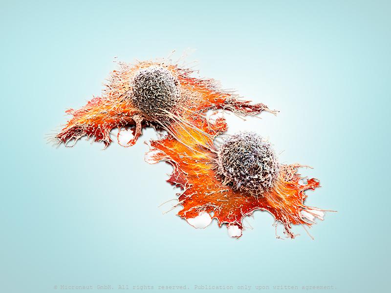
Cellular Islands (H. sapiens)
Tumors contain a vastly complicated cellular network that relies on local communication to execute malignant programs. The molecular cues that are involved in cell-cell adhesion orchestrate large-scale tumor behaviors such as proliferation and invasion. We have recently begun to understand that many tumors contain a high degree of cellular heterogeneity and are organized in a cellular hierarchy, with a cancer stem cell (CSC) population identified at the apex in multiple cancer types.

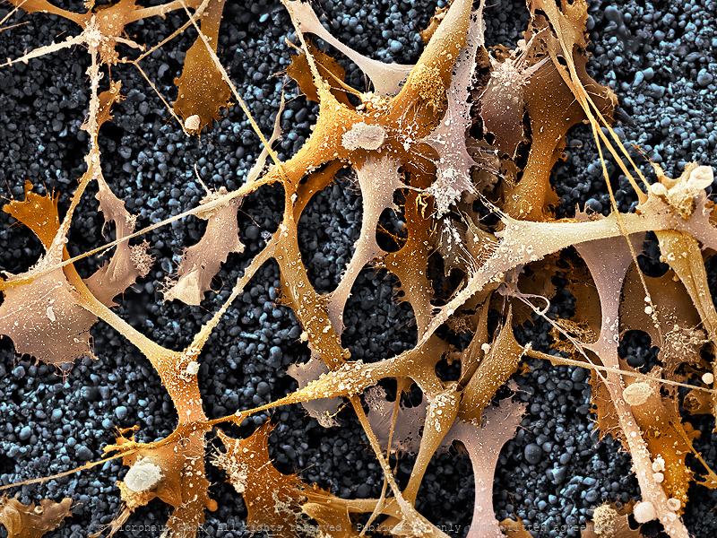
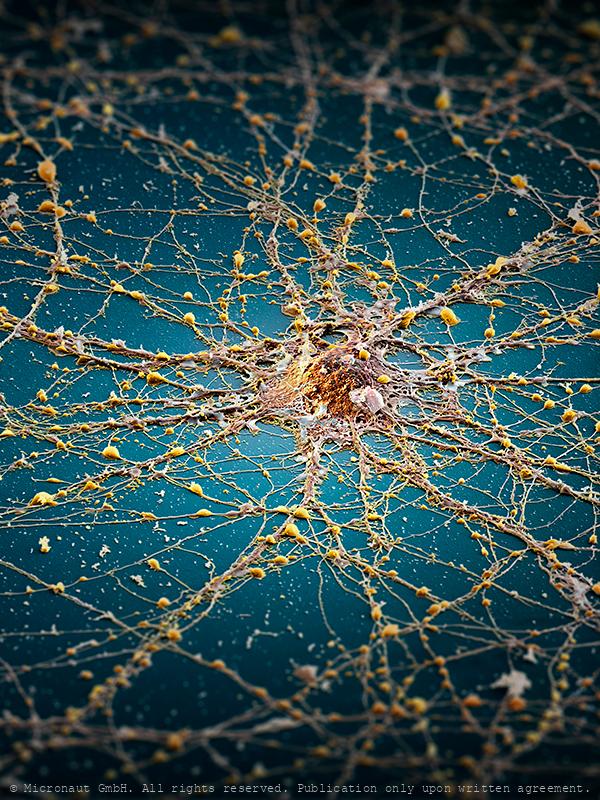
The Neuronal Megacity (Brain cell)
My concept concerning this picture was to compare a nerve cell to an island, or megacity, indicating the complex architecture of the brain. In detail, an infinite number of cellular extensions and microscopical structures unravels (capacity & complexity of the brain), whereas in the overview you can see a confusing tangle of connections that lead towards, or away, or around the center point, like the road network in a megacity. The edges of the picture are deliberately darkened and blurred, indicating there is a lot to be discovered and learned in this area of modern scientific research. Last but not least - the blue “sea” stands for Hawaii / Sophie H, to whom this picture is dedicated. Artwork created by Martin Oeggerli (Micronaut). The picture is based on scanning-electron-microscopy technology, and all colors were manually added to visualize the complex network as formed by neuronal cells in vitro.
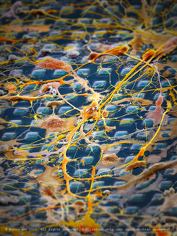
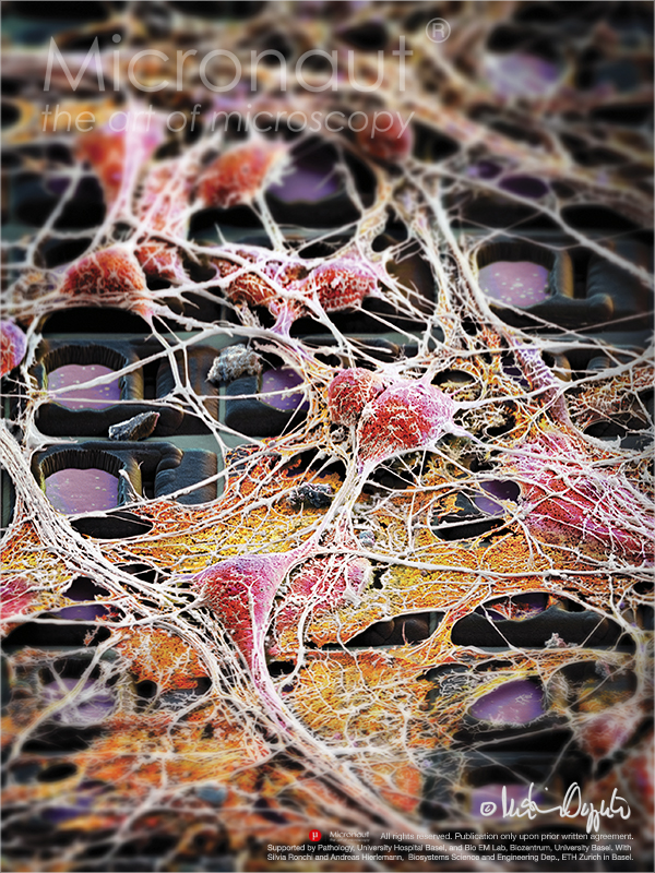
Exploring Cellular Interactions with a High-Density Microelectro
This is a CMOS-based sensor-system designed to analyse neuronal signalling, drug discovery and cell assessment. It is equipped with 26,400 electrodes and developed to function within cell-culture incubators. On top of the array, neuronal cells have been planted to explore their interactions. Every human thought starts with a signal traveling from one neuron to another in the brain. Yet we know relatively little about how these connections form and grow into the highly ordered neuronal circuits, thereby connecting billions of cells in the brain of a human adult. Understanding how neural circuits are formed is one of the fundamental questions in neuroscience. To study the growth characteristics and signalling, scientists a semiconductor chip on which they can grow and analyze cells in realtime. This artwork was created by Martin Oeggerli (Micronaut). The picture is based on scanning-electron-microscopy technology. Colors are manually added by the artist in post-production to highlight the delicate interactions between brain cells.
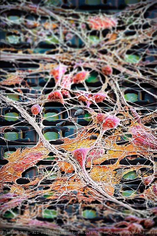
Brain Cells on a Chip (blue)
This work shows brain cells growing on a futuristic sensor-system, designed to analyse neuronal signalling. Every human thought starts with a signal traveling from one neuron to another in the brain. Yet we know relatively little about how these connections form and grow into the highly ordered neuronal circuits, thereby connecting billions of cells in the brain of a human adult. Understanding how neural circuits are formed is one of the fundamental questions in neuroscience. To study the growth characteristics and signalling, scientists have grown neuronal cells on semiconductor chip. This artwork was created by Martin Oeggerli (Micronaut). The picture is based on scanning-electron-microscopy technology. Colors are manually added by the artist in post-production to highlight the delicate interactions between brain cells.
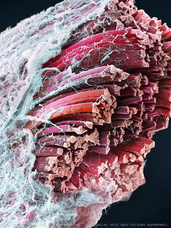
Skeletal muscle fibers (Mammalia)
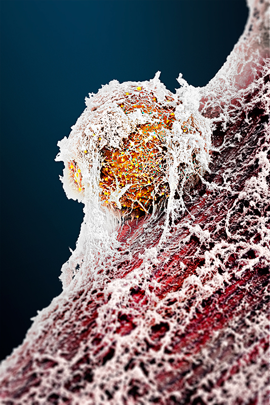
Satellite Cell (Mus musculus) - The Stem Cell that Regenerates t
2021 marks the 60th anniversary of the discovery of the "satellite cell" by Alexander Mauro by using the electron microscope and it seems an opportune moment for two researchers from the University Basel to team up with Micronaut and present a state of the art picture of a muscular stem cell. To produce this picture, mouse muscles were digested and cultured by Sedat Dilbaz and Gesa Santos in the lab of Prof. Christoph Handschin, at the Biozentrum, University Basel. Once satellite cells got activated, the sample was carefully fixed and a scanning electron microscope (SEM) was used to capture black-and-white SEM scans. After this, photorealistic colors were added in the digital darkroom by the artist. The basic unit of skeletal muscle is the myofiber: a syncytial cell packed with myofibrils, containing the sarcomeres that generate force by contraction. In vertebrates, each myofibre is controlled by many (usually hundreds) of myonuclei, which in mammals are considered post mitotic. During growth, new myonuclei are supplied by muscle satellite cells: resident stem cells located on the surface of a myofibre. In mature muscles, satellite cells become quiescent ("dormant"), but remain able to provide myoblasts for muscle hypertrophy and repair if needed. That muscle is capable of regeneration was first shown in the 1860s, but almost a century elapsed before the satellite cell was discovered. Electron microscopic studies by Alexander Mauro identified the satellite cells wedged between the plasma membrane of the muscle fibre and basement membrane and revealed that they almost completely lack cytoplasm. While nowadays, the muscle satellite cell is widely accepted as the resident stem cell of skeletal muscle, supplying myoblasts for growth, homeostasis and repair, less is known about how Satellite cells remain quiescent, or how they are activated. The understanding of the complex cellular and molecular interactions of satellite cells with their dynamic microenvironment rem
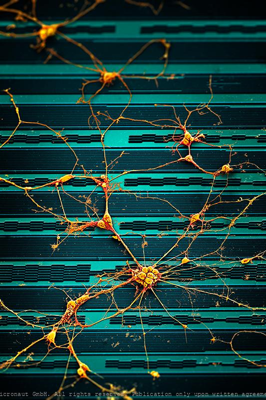
Signal Processing Analysis - Brain Cells Growing on a Microelect
Preparations of electro-active cells, predominantly brain cells, are placed directly atop fully processed microelectronics chips carrying thousands of electrodes. The chips are used to address fundamental questions in neuroscience and medicine. Artwork created by Martin Oeggerli (Micronaut). The picture is based on scanning-electron-microscopy technology, and all colors are manually added in post-production to visualize the research field of Electrophysiology and Neuroscience. We pursue an extracellular, bioelectronic approach to electrophysiology, which relies on the close juxtaposition of electrogenic cells (cells that produce electrical activity) and tissues with state-of-the-art integrated electronic systems. The cell preparations (dissociated cells, tissue slices) are placed directly atop fully processed microelectronics chips carrying thousands of electrodes and featuring CMOS circuitry. The bio-electronic interface consists of noble-metal electrodes. Prospective fields of applications of our technology include, besides fundamental neuroscience research, pharmascreening, the investigation of mechanisms of neurodegenerative diseases, or the development of aural and visual prostheses. Electrophysiology is the study of the electrical properties of biological cells and tissues. It involves measurements of voltage changes or electric currents on a wide variety of scales from single ion channels to whole organs like the heart. In neuroscience, it includes measurements of the electrical activity of neurons and, particularly, action potential activity. Neuroscience is the scientific study of molecular, cellular, developmental, structural, functional, evolutionary, computational, and medical aspects of the nervous system. It is an interdisciplinary science that includes aspects of biology, chemistry, computer science, engineering, mathematics and medicine.
Neuronal Highways - Brain Cells on a Chip
Preparations of electro-active cells, predominantly brain cells, are placed directly atop fully processed microelectronics chips carrying thousands of electrodes. The chips are used to address fundamental questions in neuroscience and medicine. Artwork created by Martin Oeggerli (Micronaut). The picture is based on scanning-electron-microscopy technology, and all colors are manually added in post-production to visualize the research field of Electrophysiology and Neuroscience. We pursue an extracellular, bioelectronic approach to electrophysiology, which relies on the close juxtaposition of electrogenic cells (cells that produce electrical activity) and tissues with state-of-the-art integrated electronic systems. The cell preparations (dissociated cells, tissue slices) are placed directly atop fully processed microelectronics chips carrying thousands of electrodes and featuring CMOS circuitry. The bio-electronic interface consists of noble-metal electrodes. Prospective fields of applications of our technology include, besides fundamental neuroscience research, pharmascreening, the investigation of mechanisms of neurodegenerative diseases, or the development of aural and visual prostheses. Electrophysiology is the study of the electrical properties of biological cells and tissues. It involves measurements of voltage changes or electric currents on a wide variety of scales from single ion channels to whole organs like the heart. In neuroscience, it includes measurements of the electrical activity of neurons and, particularly, action potential activity. Neuroscience is the scientific study of molecular, cellular, developmental, structural, functional, evolutionary, computational, and medical aspects of the nervous system. It is an interdisciplinary science that includes aspects of biology, chemistry, computer science, engineering, mathematics and medicine.
Nanomechanical Breast Cancer Identitfication
Tumor mechanobiology is an unresolved aspect of cancer progression. How the mechanical properties of cells evolve from a healthy stage to malignancy and manifest themselves in tissue has been documented and is well described in the literature but remains only poorly understood. Here, healthy ductal epithelium cells and benign lesions can be seen in the lower part and background area of the picture. In the central part, the nanomechanical characteristics of a malignous cancer cell is analysed with the tip of an Atomic Force Microscope (AFM). While a cell usually expresses numerous microvilli on its surface, invasively growing and thus more aggressive (malignant) tumor cells possess less microvilli and can be detected with AFM due to their soft stiffness profile. Detecting malignant cancer cells according to their nanomechanical properties could greatly facilitate and improve the diagnosis and treatment of e.g. breast cancer in the future.
Immortalized human cells
This hand colored scanning-electron-micrograph shows Hep-2 (ATCC CCL-23) immortalized carcinoma cells. Human epithelial type 2 (HEp-2) cells, considered to originate from a human laryngeal carcinoma, allow recognition of over 30 different nuclear and cytoplasmic patterns that are given by upwards of 50 different autoantibodies are associated with various autoimmune conditions. Hep-2 are adherent epithelial cells growing in aggregates and as single cells. Originally, Hep-2 was derived from a human larynx carcinoma of a 56-year-old black patient. However, a genetic analysis reavealed that Hep-2 is indistinguishable from HeLa (human cervix carcinoma), which means that the cell line must have been contaminated with HeLa. Nevertheless, Hep-2 is commonly used in the field of oncology and e.g. provides a particularly useful system for the study of measles, because of its growth characteristics in media free human serum.
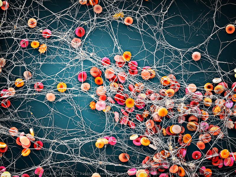
"My blood, sweat and Tears" (H. sapiens)
"My Blood, sweat, and tears have gone into this work of mine.” says Martin Oeggerli. The artist is using a scanning electron microscope to freeze time and set focus on his own blood. The final picture reveals over a hundred erythrocytes entangled in a delicate network of fibrin. Oeggerli slowly breathes life into his picture by painstakingly selecting different structures with different colors, layer upon layer on his laptop. The process allows him to set focus on the mysterious, ultra-fast and invisibly small cascade of blood clotting.Approximately 2.4 million new red blood cells (or erythrocytes) are produced per second in human adults. They are the most common type of cells in the blood and represent the vertebrate body's principal means of delivering oxygen from the lungs or gills to body tissues. Folllowing an injury, blood clotting produces a fibrin network to close the wound. Induced blood clotting can also lead to arterial embolism and result in cerebrovascular accidents, including strokes.
The Plasticity of a Tumor
Artistic coloration of cells of human KRAS-mutated pulmonary adenocarcinoma. The image shows morphologically different cells, as present within the primary lung tumor. Except some granulocytes and blood cells (lymphocytes and erythrocytes; red cells shown in the central part of the image) only tumorigenic cells are shown. The image was produced using scanning electron microscopy in combination with selectively assigning different colors to different structures. By creating a 3D-like photorealistic appearence, the artist aims to expose the complex nature and morphologic plasticity of tumor cells present in a primary tumor.
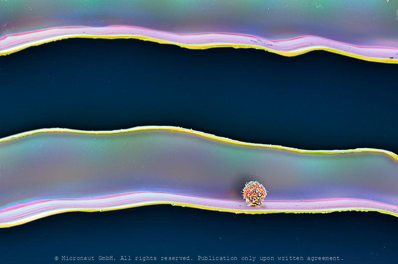
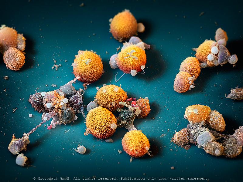
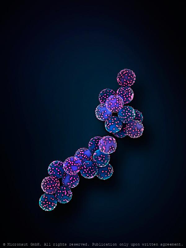
Circulating tumor cell cluster on microfluidic device
Artistic coloration of a human circulating tumor cell cluster, isolated from the blood of a patient with cancer using a microfluidic device, by Martin Oeggerli (Micronaut). Circulating tumor cells are cancer cells that detach from the primary tumor and enter the blood circulation, on their way to initiate a metastasis at a distant site. When traveling in the blood as clusters, circulating tumor cells are endowed with an extraordinary potential to efficiently initiate a metastasis. While very rare compared to blood cells in the bloodstream (1 circulating tumor cell per billion blood cells), capturing of circulating tumor cell clusters is now possible through the use of specialized microfluidic technologies, thus enabling scientists to study their biology and vulnerabilities.
Virus Imprinted Nanomaterial Particles
Artificial virus recognition nanomaterials- Scanning electron micrograph of virus imprinted particles - VIPs - created via a surface initiated bottom-up synthetic approach using viruses as templates. The blue particles (approx. 400 nm) represent the VIPs while the "pink" dots represent the trapped virus.
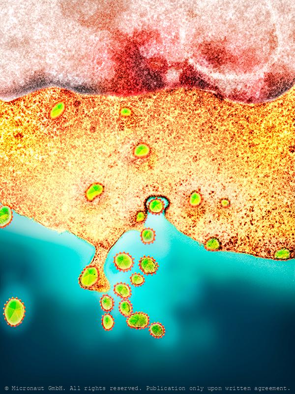
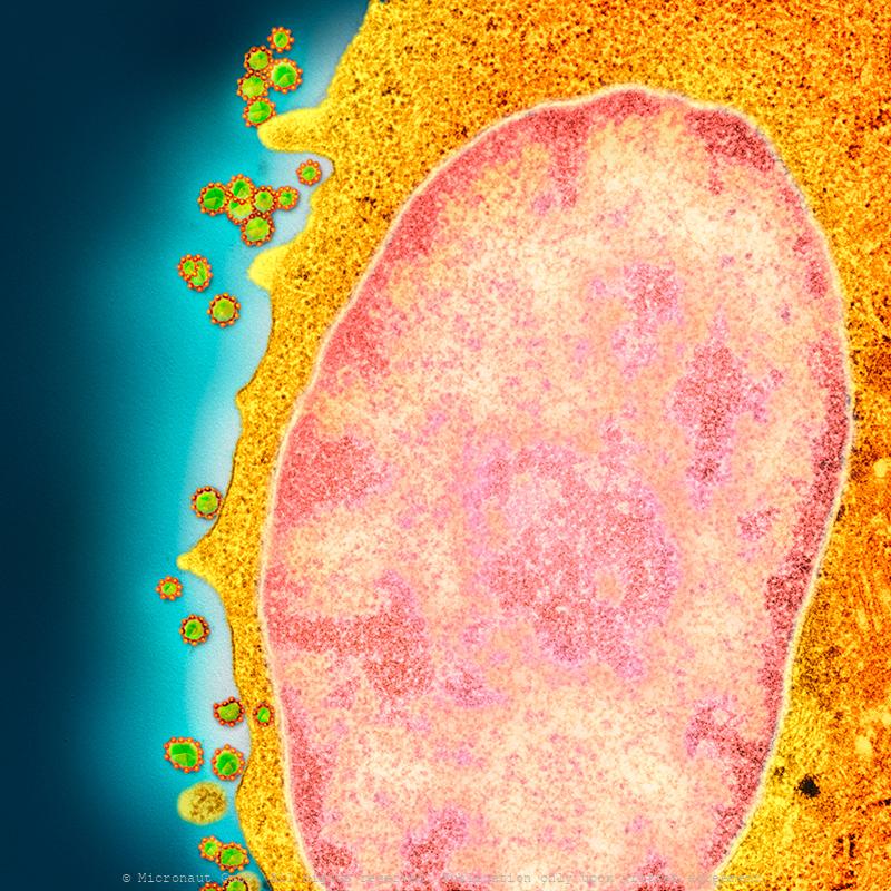
HIV (Nr. 2)
HIV viruses infecting a cell. Transmission electron micrograph (TEM) of a section through human immunodeficiency virus (HIV) particles (virions, round) on the surface of a cultured cell. HIV attacks CD4+ T-lymphocytes (specialised white blood cells), which are crucial in the body's immune system. It enters the cell and makes many copies of itself, which then destroy the cell as they emerge through its membrane. This severely weakens the immune system, causing AIDS (acquired immunodeficiency syndrome).
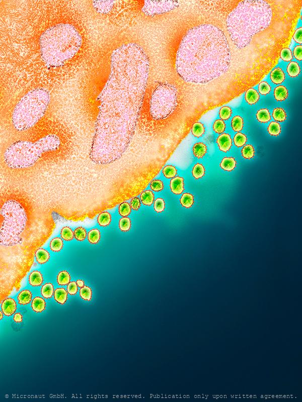
HIV (Nr. 1)
HIV viruses infecting a cell. Transmission electron micrograph (TEM) of a section through human immunodeficiency virus (HIV) particles (virions, round) on the surface of a cultured cell. HIV attacks CD4+ T-lymphocytes (specialised white blood cells), which are crucial in the body's immune system. It enters the cell and makes many copies of itself, which then destroy the cell as they emerge through its membrane. This severely weakens the immune system, causing AIDS (acquired immunodeficiency syndrome).
HIV (Nr. 3)
HIV viruses infecting a cell. Transmission electron micrograph (TEM) of a section through human immunodeficiency virus (HIV) particles (virions, round) on the surface of a cultured cell. HIV attacks CD4+ T-lymphocytes (specialised white blood cells), which are crucial in the body's immune system. It enters the cell and makes many copies of itself, which then destroy the cell as they emerge through its membrane. This severely weakens the immune system, causing AIDS (acquired immunodeficiency syndrome).
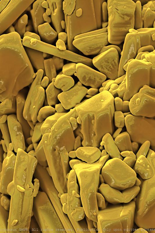
Doxycycline - Antibiotics
Antibiotics (Doxycycline)
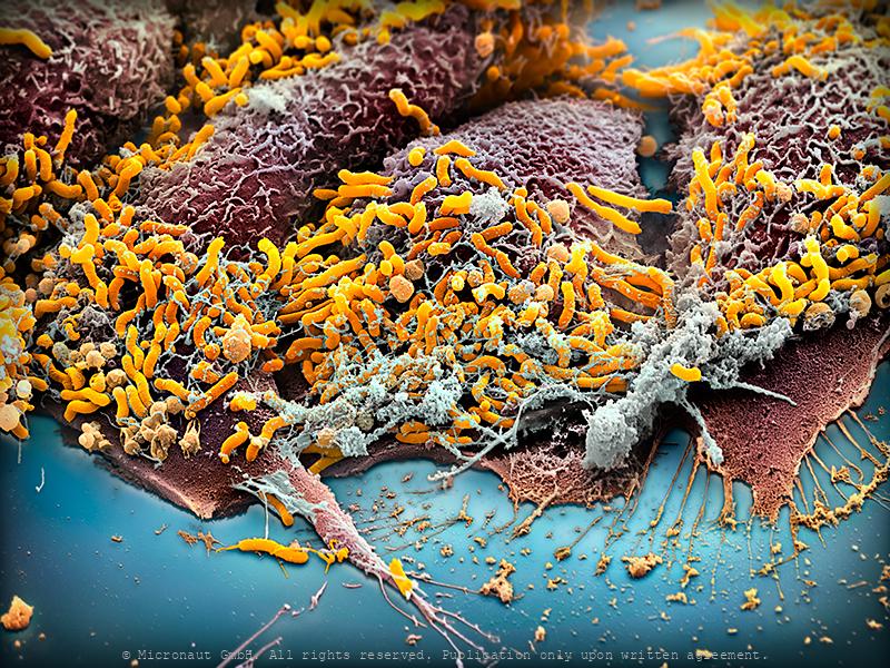
Stomach cells infection - Helicobacter pylori
In vitro cultured stomach cancer cells (red/brown), infected with Helicobacter pylori (yellow). Magenepithelzellen wurden in vitro gezüchtet und experimentell infiziert mit Helicobacter pylori; die Bakterien sind in gelb zu sehen, die Wirtszellen sind rot-braun. In vitro cultured stomach cancer cells (brown), infected with Helicobacter pylori (yellow). Helicobacter pylori, a common bacterium that lives in the stomach lining, increases the risk of stomach cancer and peptic ulcers. But over time H. pylori can reduce stomach acid and acid reflux, which may help fend off esophageal cancer. The microbe also appears to help protect us from allergies and asthma. Some scientists suspect that the dramatic increase in those conditions in the industrialized world could be related to the decreasing frequency of H. pylori in our stomachs, which is partly due to high doses of antibiotics in childhood.
The Magic Bullet - Cnidocyte
The name Cnidaria comes from the Greek word "cnidos", which means stinging nettle. Invisibly small, the cnidocyst which are responsible for the explosive pain caused by jellyfish, remains a mysterious weapon until today. Cnidarians are a varied group of animals including corals, sea anemones, sea pens, sea pansies, jellyfishes, and hydroids. They range among the most beautiful of all marine invertebrates. Many species have a flower-like look and are often considered delicate and soft. But they possess a well-concealed yet potent arsenal of harpoon-like stinging cells. Stings from several species can be very painful, and in some cases, even fatal. Box jellyfish and sea wasps represent the biggest “stinging” threat to divers and swimmers. Portuguese men-of-war are colonial hydroids and their tentacles laced with potent stinging cells are hidden underneath the purple-to-blue float bobbing on the surface. In some specimens the tentacles can reach 20 feet. The virulent stinging cells are easily capable of paralyzing any pelagic fish and in humans can cause extreme pain requiring medical attention. But how does the toxic weapon look like? Cnidocytes are small compartments that house a mini needle-like stinger. When an outside force triggers the stinger, the cell opens, letting ocean water rush in. This causes the stinger to shoot out into what triggered the action; once it’s there, venom is released. All of this happens within a millionth of a second. This picture shows a discharged toxic cnidocyte (i.e. one that has been fired) of a freshwater polyp. It was created with the scanning-electron-microscope by science photographer Martin Oeggerli. Based on the original SEM scan, he colored the picture layer-by-layer, to uncover the delicate structures and shed light on the anatomy of this miniature injection tool.
Viral infection - The Parasites of Parasites of Parasites... Nr.1
A bacteriophage is a virus that infects and replicates within a bacterium. It is composed of proteins that encapsulate a DNA or RNA genome. Bacteriophages ('phages') have relatively simple structures and their genomes encodes something between four and several hundred genes. To replicate, phages inject their genome into the cytoplasm of a bacteria. Bacteriophages are among the most common and diverse entities in the biosphere. They are widely distributed in locations populated by bacterial hosts, such as soil or the intestines of animals. One of the densest natural sources for phages and other viruses is sea water. This hand colored scanning-electron-micrograph shows bacteria which are heavily infected by bacteriophages. At the lower right hand side, a bacterial cell has been broken open (lysed) and destroyed. As soon as the cell is destroyed, the phage progeny can find new hosts to infect.
Viral infection - The Parasites of Parasites of Parasites... Nr.1
A bacteriophage is a virus that infects and replicates within a bacterium. It is composed of proteins that encapsulate a DNA or RNA genome. Bacteriophages ('phages') have relatively simple structures and their genomes encodes something between four and several hundred genes. To replicate, phages inject their genome into the cytoplasm of a bacteria. Bacteriophages are among the most common and diverse entities in the biosphere. They are widely distributed in locations populated by bacterial hosts, such as soil or the intestines of animals. One of the densest natural sources for phages and other viruses is sea water. This hand colored scanning-electron-micrograph shows bacteria which are heavily infected by bacteriophages.
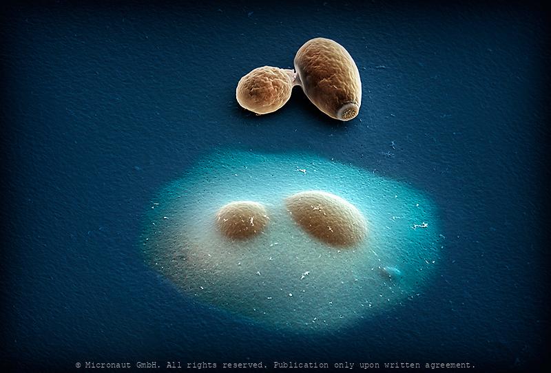
Encapsulated vs. normal yeast cells
Budding yeast (Saccharomyces cerevisiae); hand-colored scanning-electron-micrograph by Micronaut (Martin Oeggerli). Modification of cell surfaces with synthetic hydrogels is a promising approach for controlling cell behaviour. Here, Vanella and colleagues developed a genetically controlled system that enables cells to auto-encapsulate inside protective hydrogel capsules. The image shows an encapsulated budding yeast cell directly adjacent to a non-encapsulated one. This system links inducible gene expression to a synthetic capsule that protects target cells against lysis and mediates communication with the extracellular space through a permeable hydrogel.
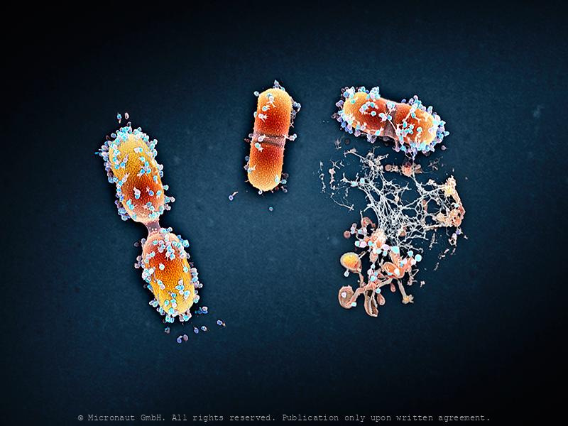
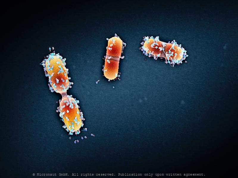
E. coli on an LP -(CRISPR-Cas Mediated Genome Editing)
This hand-colored scanning-electron-micrograph (SEM) shows cells of the human gut bacterium Escherichia coli (E. coli) on a phonograph record of Ludwig van Beethoven’s Moonlight Sonata (Sonata No 14 in C). E. coli has been engineered to express the molecular components of another bacterium’s CRISPR-Cas system - an adaptive bacterial immune system that enables it to write a permanent record of the RNAs within the cell into its own genome where it is safely stored. Much like the Beethoven’s composition, this record is preserved over many generations. Scientist can retrieve it at any time to reveal the mysteries of the bacterium’s transcriptional history.
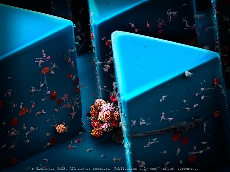
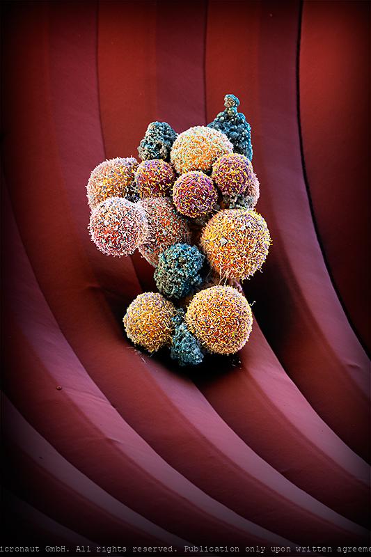
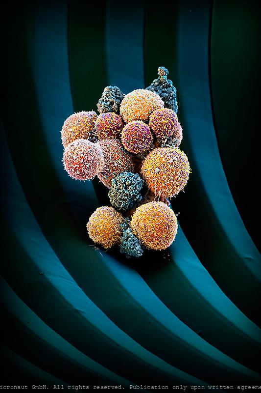
Circulating Tumor Cells, Nr.1
Cancer cells are carried through the bloodstream either as single circulating tumor cells (CTCs) or as multicellular groupings (CTC clusters). In contrast to relatively small single CTCs, the number and dimension of CTC clusters favours the formation of new matastasis sites. This artistically colored scanning-electron-micrograph (SEM) illustrates the new microchip technology which is used to capture and analyse human CTC clusters. By analysing a blood sample of a breast cancer patient with a microfluidic device (blue), blood cells and CTCs can be enrichted and picked and prepared for further examination from the smooth surface of the chips triangle-microstructures. At the center of the image you can see a typical CTC cluster, containing 9-12 tumor cells. Circulating tumor cells are believed to exist in most - if not all - tumor types. Once the process of matastasis has started, the number of circulating tumor cells is continuously increased: i.e. in a 20ml blood sample up to 100 billion blood-cells and 10 CTCs, or CTC clusters can be found. It is important to analyse the morphology and biology of CTC clusters: How do they form? What binds single cells in a CTC cluster? How can CTC clusters speed up metastasis formation? The long-term goal of such research projects is to treat and eliminate CTC clusters in patients, thereby preventing primary tumors to metastasize. -- Klumpen aus zirkulierenden Tumorzellen (CTCs) einer Patientin mit metastasierendem Brustkrebs werden mit Hilfe eines selbst entwickelten Mikrofluidik-Chips (blau) isoliert. Als Vergleichsgrösse dienen rote Blutkörperchen (7.5 µm, rot). © Martin Oeggerli, Micronaut.ch
Dividing Streptococcus (beta-haem; remastered version)
dividing streptococcus sp. bacteria
Circulating tumor cell cluster on microfluidic device
Artistic coloration of a human circulating tumor cell cluster, isolated from the blood of a patient with cancer using a microfluidic device, by Martin Oeggerli (Micronaut). Circulating tumor cells are cancer cells that detach from the primary tumor and enter the blood circulation, on their way to initiate a metastasis at a distant site. When traveling in the blood as clusters, circulating tumor cells are endowed with an extraordinary potential to efficiently initiate a metastasis. While very rare compared to blood cells in the bloodstream (1 circulating tumor cell per billion blood cells), capturing of circulating tumor cell clusters is now possible through the use of specialized microfluidic technologies, thus enabling scientists to study their biology and vulnerabilities.
Cellular network
Prostate cancer (in-vitro) cell culture. LNCaP cells are a cell line of human cells commonly used in the field of oncology. LNCaP cells are androgen-sensitive and derive from a human prostate adenocarcinoma (lymph node metastasis) of a 50-year-old caucasian male patient. They are adherent epithelial cells growing in aggregates and as single cells. Prostate cancer is the most frequently diagnosed cancer in men. One major obstacle in prostate cancer research is the lack of cell lines that closely mimic human disease progression. Two hallmarks of metastatic human prostate cancer include the shift of aggressive PCa from androgen-sensitivity to an androgen insensitive state, and the preference of primary prostate cancer cells to metastasize to bone. Although the generation of androgen insensitive cell lines has been demonstrated in “classic” cell lines (DU145 and PC3), the behavior of these cells in bone metastasis does not fully mimic clinical human disease. Therefore, LNCaP sublines have been generated to develop an androgen insensitive prostate cancer cell model that more closely mimics clinical disease.
Targeting the Achilles Heel of a Tumor
Immune system (T cell) attacks a human tumor cell (non small cell lung cancer). Manually colored 3D realistic scanning electron micrograph by Martin Oeggerli / Micronaut, 2019. Immunotherapy is becoming increasingly important. “What’s revolutionary about it is that some patients may remain cured after years of treatment – even in the case of tumours that have otherwise proved resistant to therapy”, says Zippelius. In the meanwhile, University Hospital Basel has set up its own tumour board (a group of doctors with different specialities) for immunotherapy. The project is a collaboration between the Department of Medical Oncology and the Department of Thoracic Surgery at University Hospital Basel, the Netherlands Cancer Institute in Amsterdam as well as the Department of Pathology at the Cantonal Hospital Baselland in Liestal and the Roche Innovation Center Basel.
Smallpox virus (Vaccinia sp.)
Conquered Killer – Smallpox ranks among the most devastating illnesses ever suffered by humankind. For centuries, it killed millions of people around the world. Thanks to global immunization programs, the deadly infectious disease was wiped out in 1979 following a successful vaccination program regarded as one of the greatest triumphs of modern medicine. Pockenviren sind die größten und bekanntesten Tierviren. Sie weisen Eigenschaften auf, die denen primitiver Zellen ähneln. Jedoch sind die Pockenviren, wie alle Viren, nicht in der Lage, Stoffwechsel zu betreiben, und sind daher in Bezug auf die Proteinbiosynthese vom Wirt abhängig. Diese Viren sind hinsichtlich DNA-Replikation ungewöhnlich: Eine mit dem Pockenvirus infizierte Zelle weist eine DNA-Synthese außerhalb des Kernes auf, was sonst nur in intrazellulären Organellen wie Mitochondrien (und bei Pflanzen in Chloroplasten) oder bei einer Infektion mit dem Hepatitis-B-Virus vorkommt. Bei den echten Pocken (Variola vera und Variola major) liegt die Sterblichkeit bei 10-90%. Die Übertragung erfolgt über Tröpfcheninfektion, zB bei Husten. Beim Krankheitsverlauf kommt es zu auffälligen Hautveränderungen (Pustel, Eiterbläschen). Für die Impfung werden Kuhpocken (Vaccinia Viren) verwendet. Diese sind im Bild zu sehen.
human blood cells during coagulation process
human blood cells during coagulation process
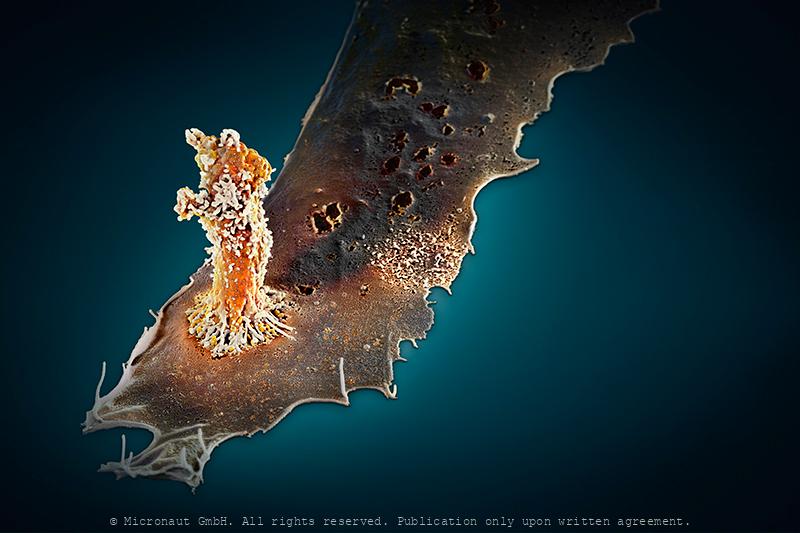
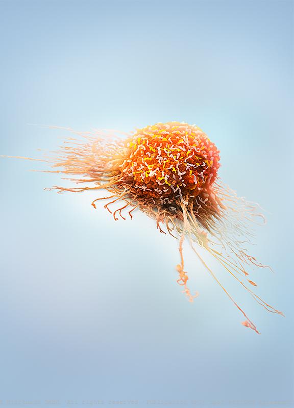
Invasively Growing Cancer Cell
Invasively growing human cancer cell, in vitro. Cancer cells develop the ability to move themselves, sooner or later. The picture shows an invasively growing tumor cell, which is pulling itself around with the help of its appendages. In comparison to a healthy cell its extensions are smaller and because of the quicker assembly and disassembly of the appendixes, it is able to move faster. -- Die Transformation einer gesunden Zelle in eine Tumorzelle ist ein komplexer Vorgang und bedingt eine ganze Reihe verschiedener genetischer Veränderungen. Tumorzellen entwickeln dabei früher oder später die Fähigkeit sich fortzubewegen und Metastasen zu bilden. Das Bild zeigt eine invasive Prostatakarzinomzelle, welche häufig Metastasen im Knochenmark bilden. Mit Hilfe der Fortsätze zieht sich die Zelle über die Unterlage. Im Vergleich zu einer gesunden Zelle bildet diese invasive Tumorzelle deutlich kleinere Fortsätze aus und ist dank schnellerem Auf- und Abbau (der Fortsätze) in der Lage, sich wesentlich schneller fortzubewegen.
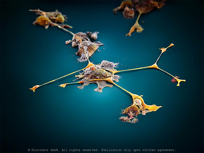
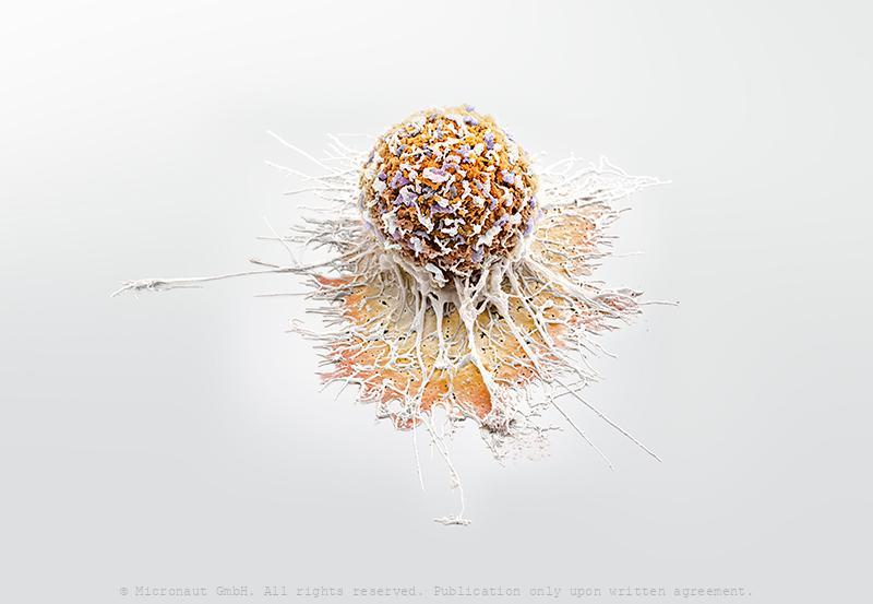
Cancer Cell Extensions
Invasively growing prostate cancer (in-vitro) cell culture. Prostate cancer is a form of cancer that develops in the prostate, a gland in the male reproductive system. Most prostate cancers are slow growing and are diagnosed late in life (after 75 yrs). However, aggressive prostate cancer cells may arise and spread (metastasize) to other parts of the body, particularly into bones and lymph nodes. From the genetical background, no single gene is responsible for prostate cancer. In contrast, many different genes are implicated, including oncogenes and tumor suppressor genes. Mutations in BRCA1 and BRCA2, which are also important risk factors for ovarian cancer and breast cancer in women, are implicated in prostate cancer as well. Other oncogenes that play an important role in prostate cancer include: the Hereditary Prostate cancer gene 1 (HPC1), the vitamin D receptor, gene fusions with the TMPRSS2 gene family - and especially the androgen receptor which is most often involved in the development and progression of the disease.
Clonal evolution versus cancer stem cell theory
In cancer research two fundamentally different mechanisms explaining tumor progression currently split the positions: Clonal evolution versus cancer stem cells. To develop more effective cancer therapies it will be critical to determine which theory holds true. If most cells can proliferatie and metastasize, then virtually all cells must be eradicated to cure the disease, whereas the specific elimination of cancer stem cells would be sufficient, if the theory of clonal evolution is a myth...
The Neuronal Megacity (Brain cell)
My concept concerning this picture was to compare a nerve cell to an island, or megacity, indicating the complex architecture of the brain. In detail, an infinite number of cellular extensions and microscopical structures unravels (capacity & complexity of the brain), whereas in the overview you can see a confusing tangle of connections that lead towards, or away, or around the center point, like the road network in a megacity. The edges of the picture are deliberately darkened and blurred, indicating there is a lot to be discovered and learned in this area of modern scientific research. Last but not least - the blue “sea” stands for Hawaii / Sophie H, to whom this picture is dedicated. Artwork created by Martin Oeggerli (Micronaut). The picture is based on scanning-electron-microscopy technology, and all colors were manually added to visualize the complex network as formed by neuronal cells in vitro.